

 PDF
PDF ePub
ePub Citation
 Print
Print


Abstract
Purpose:
Incidence of radiographic contrast media extravasation has increased owing to the escalating availability of contrast enhanced imaging. Potential complications of extravasation include localized swelling, itching sensation, hypesthesia, erythema, limitation of finger movement, compartment syndrome, skin sloughing, and necrosis. We describe clinical characteristics and treatment of computed tomography contrast media extravasation.
Methods:
A retrospective chart review was performed on 26 consulted patients experiencing contrast extravasation, between January 2005 and December 2011. Age, signs, symptoms, types of contrast administered, volume of extravasation, treatment and patient outcomes were documented and evaluated, retrospectively.
Results:
Extravasation of less than 100 mL occurred in 85%. Immediate surgical therapy was necessary in 23% of cases. There were no postoperative complications and it rendered excellent cosmetic outcomes. In 77% of cases, conservative management was recommended, such as elevation and immobilization of extremity, application of ice pack, and careful monitoring.
REFERENCES
1. Hannon MG, Lee SK. Extravasation injuries. J Hand Surg Am. 2011; 36:2060–5.

2. Sistrom CL, Gay SB, Peffley L. Extravasation of iopamidol and iohexol during contrast-enhanced CT: report of 28 cases. Radiology. 1991; 180:707–10.
3. Federle MP, Chang PJ, Confer S, Ozgun B. Frequency and effects of extravasation of ionic and nonionic CT contrast media during rapid bolus injection. Radiology. 1998; 206:637–40.
4. Sbitany H, Koltz PF, Mays C, Girotto JA, Langstein HN. CT contrast extravasation in the upper extremity: strategies for management. Int J Surg. 2010; 8:384–6.
5. Gothlin J, Hallbook T. Skin necrosis following extravasal injection of contrast medium in phlebography. Radiologe. 1971; 11:161–5.
6. Seiler JG III, Olvey SP. Compartment syndromes of the hand and forearm. J Am Soc Surg Hand. 2003; 3:184–98.
7. Kumar RJ, Pegg SP, Kimble RM. Management of extravasation injuries. ANZ J Surg. 2001; 71:285–9.
8. Schaverien MV, Evison D, McCulley SJ. Management of large volume CT contrast medium extravasation injury: technical refinement and literature review. J Plast Reconstr Aesthet Surg. 2008; 61:562–5.
9. Tong R. Preventing extravasation injuries in neonates. Paediatr Nurs. 2007; 19:22–5.
10. Bellin MF, Jakobsen JA, Tomassin I, et al. Contrast medium extravasation injury: guidelines for prevention and management. Eur Radiol. 2002; 12:2807–12.
11. Rowlett J. Extravasation of contrast media managed with recombinant human hyaluronidase. Am J Emerg Med. 2012; 30:2102.e1–3.
12. Loth TS, Eversmann WW Jr. Extravasation injuries in the upper extremity. Clin Orthop Relat Res. 1991; (272):248–54.
13. Mubarak SJ. A practical approach to compartmental syndromes. Part II. Diagnosis. Instr Course Lect. 1983; 32:92–102.
14. Mubarak SJ, Hargens AR. Acute compartment syndromes. Surg Clin North Am. 1983; 63:539–65.
15. Whitesides TE Jr, Haney TC, Harada H, Holmes HE, Morimoto K. A simple method for tissue pressure determination. Arch Surg. 1975; 110:1311–3.
Fig. 1.
Case 1. (A) A 59-year-old female showing erythema, swelling, paresthesia, blistering and severe pain. (B, C) Intraoperative view. Fasciotomy and concomitant transverse carpal ligament release were performed. (D) Postoperative 6 months follow-up revealed excellent functional and cosmetic outcomes.
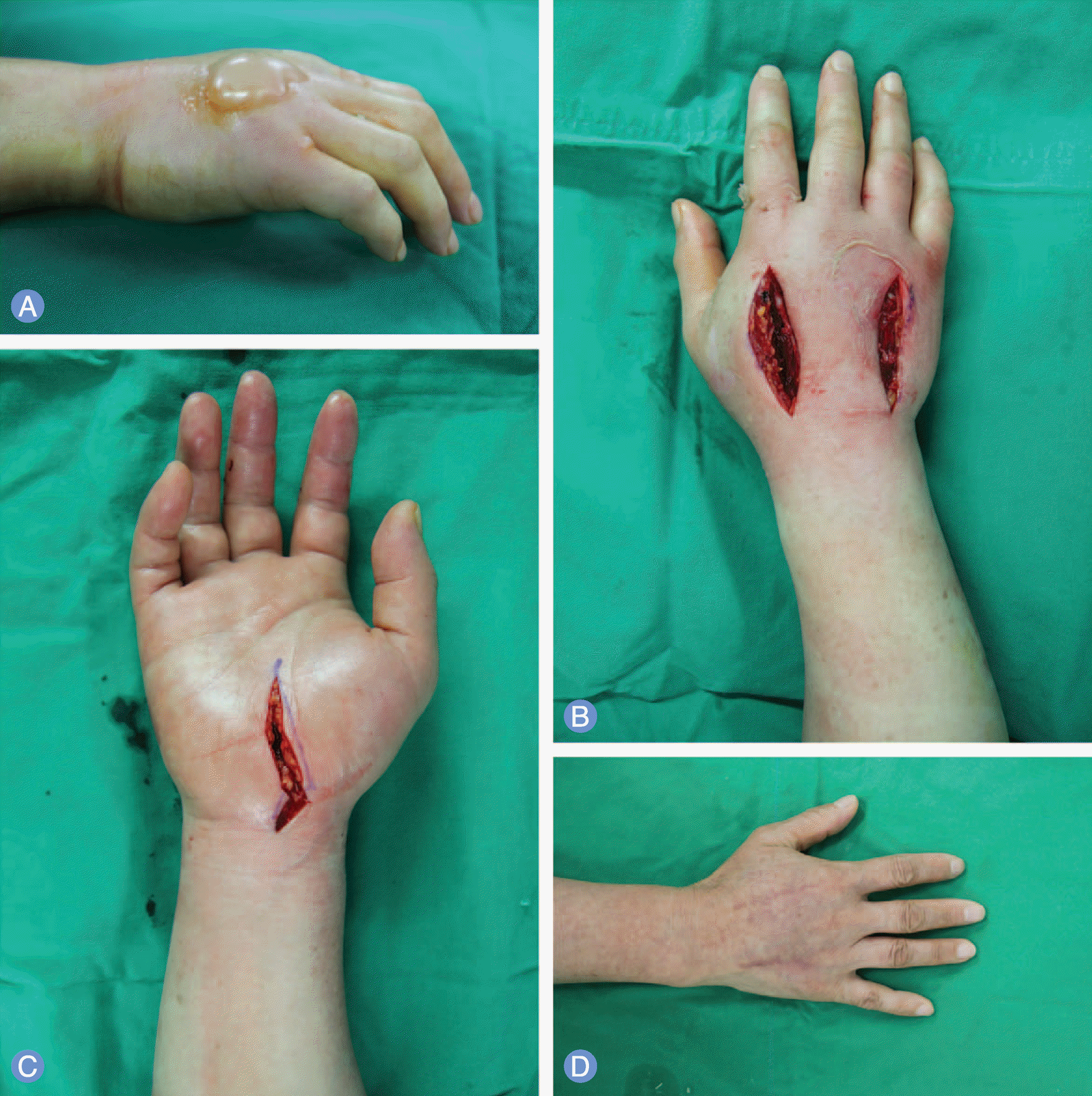
Fig. 2.
Case 2. (A) A 57-year-old female showing swelling and paresthesia. (B) Intraoperative view. An incision was made on the dorsum of the hand and saline washout was performed. (C) Postoperative 6 months follow-up revealed excellent functional and cosmetic outcomes.
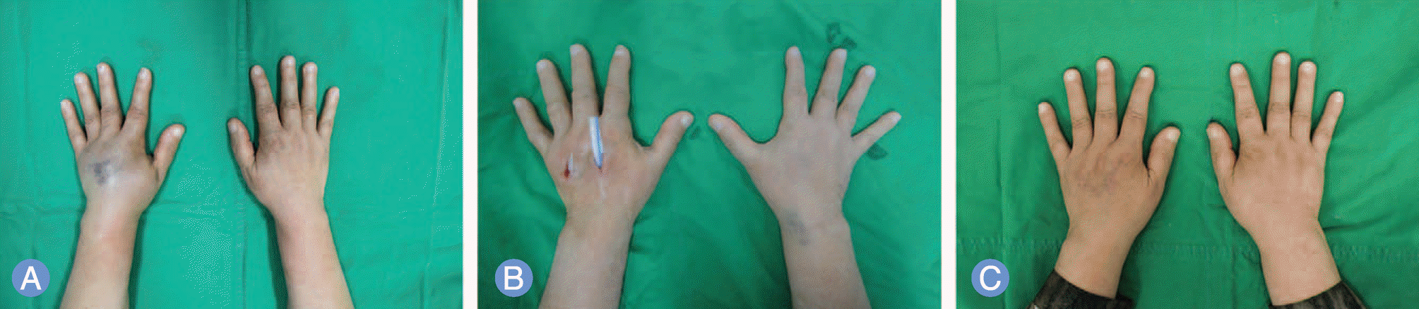
Table 1.
Summary of 26 patients
 XML Download