

 PDF
PDF ePub
ePub Citation
 Print
Print


Guyon's canal in the wrist is the passage of the ulnar nerve located at the base of the wrist and it is a structure that extends from the proximal aspect of the palmar carpal ligament to the fibrous bow of the hypothenar muscle1. Because the ulnar nerve is divided into the superficial sensory branch and the deep motor branch within Guyon's canal, the extent of motor impairment and sensory loss may vary depending on which part of Guyon's canal is damaged.
Scar formation after tissue injury is an essential part of wound healing. The injured peripheral nerve often thickens the nerve endothelium, forms scar tissue around the nerve, and scar tissue induces adhesion between the surrounding critical structures. It can then progress to chronic retraction and permanent nerve compression due to peripheral nerve compression and retraction.
In this paper, we report a case where we successfully performed a surgical technique of nerve wrapping using a silicone tube when performing neurorrhaphy in order to prevent neuropathy caused by scar tissue formation after performing nerve repair to treat a deep laceration at the area of the Guyon's canal in the wrist, and we present the case report with a review of the literature.
CASE REPORT
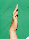
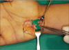
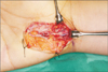
An 18-year-old female patient visited the emergency department after she fell in the bathroom and suffered a deep laceration of the right palm at the sharp part of the tile floor. The patient was discharged after debridement and primary repair. On the 9th day after the injury, she visited the hospital for outpatient treatment and complained of sensory impairment in the outer half of the fourth and fifth digits of the hand and motor function disturbance. The patient was diagnosed as having clawhand deformity at the time of the physical examination (Fig. 1). Physical examination revealed that the patient did not spread the fingers of the injured hand apart or draw them together well, and the results of Froment's sign were positive (Fig. 2). The results of simple radiographs of the hand were normal findings. Ulnar nerve injury was suspected in the electromyogram and nerve conduction study performed on the 12th day after the injury. On the 23rd day after the injury, the skin was incised along the sutured wound using general anesthesia and tourniquet, and the Guyon's canal was exposed, and it was found that the ulnar nerve and ulnar artery were completely cutted and adhered to the surrounding tissues (Fig. 3). After finding the severed ulnar nerve and artery, adhesions with the surrounding tissues were resolved, and neurorrhaphy was performed on the deep and superficial branches of the ulnar nerve. At the same time, a commercially available silicone tube (Silicone tube; Sun Medical Co., Seoul, Korea) was used to wrap the ulnar nerve in the area where there was a possibility of adhesion with surrounding tissue or compression (Fig. 4). After performing arteriorrhaphy, the wound was sutured and a short arm splint was applied for 2 weeks after surgery. Before entering operationg room, written informed consent was obtained from the patient after informing rare possibility of complications caused by implantation of foreign substances. The results of the Hand Grip Power and Dexterity test performed on the 18th postoperative day showed a significant improvement in muscle strength and motor function compared with the results of the same test on the 8th day (Table 1, 2). At 8 months postoperatively, the electromyogram showed a significant improvement compared to the preoperative level. When the Jebsen-Taylor Hand Function Test was performed 9 months after surgery, the results were similar to the normal values in most of the items (Table 3). The patient did not develop any complications including discomfort, which is most common complication of silicone tube during the follow-up period of 2 years and showed continuous improvement of motor and sensory function through rehabilitation.
DISCUSSION
Because the ulnar nerve is located relatively close to the skin when passing through the elbows and wrists, it is very likely to be damaged in these areas. Shea and McClain2 classified the injuries of the ulnar nerve in the wrist into three categories according to the injury location. Type 1 is sensory and motor deficit due to the injury of the area proximal to or in the canal of Guyon, Type 2 is motor impairment due to the injury of the deep branch of the ulnar nerve, and Type 3 is sensory loss due to the injury of the distal part of the ulnar canal where the superficial branch of the ulnar nerve is located.
Perineural scar formation and adhesion can be induced by traumatic injury, hemorrhage of the surgical site, chronic inflammation, or a simple surgical operation. Scar formation after neurorrhaphy is considered to be inevitable3. After undergoing decompression because of carpal tunnel syndrome, 20% of the patients reported recurrence of the symptom because of scar tissue formation, and it has been reported that adhesion of the nerve and surrounding tissue recurs in most patients even after secondary neurolysis4.
If perineural scar formation occurs, the nerve is compressed and the compressed nerve causes irreversible damage as well as ischemic changes, resulting in symptoms such as sensory deficit, muscle atrophy, functional disability, and chronic pain. In addition, nerve regeneration is interfered by scar tissue itself5.
For this reason, previous studies continued to investigate various surgical techniques and pharmacological treatments to minimize scar formation around the nerve after neurorrhaphy. In order to minimize scar formation, methods such as microsurgery, endoscopy, nerve transposition, and fat graft have been performed. In some cases, the operation site was irradiated with radiation after surgery, and methods using laser irradiation, fibrin glue, and the like have also been used as an attempt to reduce foreign body reaction by the suture material itself67. In addition, since the use of triamcinolone acetonide and cishydroxyproline for suppression of scar formation after neurosurgical operations in the 1960s, various medicines such as amniotic fluid and hyaluronic acid have been used with the expectation that they would help suppress scar formation after neurorrhaphy89. However, previous studies on the surgical procedures and pharmacological treatments described above did not provide a consistent and satisfactory result, and the ultimate treatment for prevention of scar formation after neuronal surgery has not been presented yet.
Nerve wrapping was derived from the concept of suppressing over-activated fibroblast reaction after a neurosurgical operation by placing a biologic barrier consisting of fascia, fat, and vein graft around the damaged nerve. Actually, nerve wrapping using the saphenous vein has been performed in cases of chronic median nerve compression. Recently, silicone tubing has been successfully used for peripheral nerve repair and major complications have not been reported10.
In respect of the case described in this paper, we performed neurorrhaphy for the patient whose ulnar nerve injury was not detected immediately but found later, and additionally tried the surgical technique of nerve wrapping with a commercial silicone tube, employing the concept of nerve wrapping used to prevent neuropathy caused by scar formation after neurorrhaphy. After surgical treatment, motor and sensory deficits due to ulnar nerve injury were significantly improved, and the patient recovered without complications until 2 years postoperatively. Continuous follow-up is needed to confirm that there are no complications, including nerve compression and foreign body reactions. However, wrapping the nerve with a silicone tube is expected to prevent the occurrence of scar-induced neuropathy not only in patients who have undergone nerve repair because of ulnar nerve injury as in the case reported in this study but also in patients undergoing surgical treatment such as nerve decompression because of Guyon's canal syndrome due to other causes.


 XML Download