

 PDF
PDF ePub
ePub Citation
 Print
Print


Introduction
Dysphagia is common in stroke patients and its incidence is various by report from 51% to 73%.1,2 According to a prior study, in a clinical dysphagia evaluation 6 months after stroke, 6~11% of the patients still suffered from symptoms of dysphagia,3,4 and when assessed with a videofluoroscopic swallowing study (VFSS), 28~50% of the patients were confirmed to have dysphagia.4
Normal swallowing is a complex and sequential process in which oral phase, pharyngeal phase and esophageal phase are in harmony, and it is known that dysphagia occurs when there is a lesion in the brain stem where the nucleus of the swallowing reflex is located.5 In general, lateral fissure (insula) cortex is considered as the cerebral swallowing center.6 Cerebral cortical infarction can cause dysphagia but it is still controversial which of the two hemispheres plays a more important role in swallowing.7 Some previous studies reported that there were different patterns of dysphagia between right and left hemisphere lesion, however, others reported there were no different patterns of dysphagia between the two.7,8,9 There are many conflicting studies on the association between the lesion location and the patterns of dysphagia, which has not been clearly identified yet.
While there were a lot of studies on the diagnosis and treatment of dysphagia in stroke patients, there were only limited studies published for to enlighten clinical factors associated with dysphagia.
Thus, we aimed to investigate the factors associated with dysphagia such as the brain lesion location, cognitive functions and the patient's characteristics (age, gender and type of stroke, etc.) via retrospective chart review.
Materials and Methods
1) Subjects
Subjects of this study were first-ever subacute stroke patients who had undergone a VFSS to screen or to evaluate for dysphagia in the Department of Rehabilitation Medicine at OO Medical Center from January 2006 to April 2012. Stroke was diagnosed only in case of cerebral infarction or hemorrhage had been verified on the basis of brain computed tomography (CT) or magnetic resonance imaging (MRI). The exclusion criteria were as follow; 1) Patients who had disease that might cause dysphagia. 2) Patients with bilateral stroke or a previous stroke history. 3) Patients without a CT or MRI results. 4) Patients whose lesion could not be classified like subarachnoid hemorrhage (SAH). 5) Patients who were unable to perform an appropriate VFSS due to their poor cooperation. 6) Cases in which the interval between the Korean version of mini-mental state examination (K-MMSE) and VFSS evaluations was longer than 2 weeks. A total of 178 patients met the inclusion criteria and were chosen for study, and their data were collected retrospectively from their medical records.
2) Methods
We reviewed the patients' medical records and test results retrospectively, and their demographic and clinical characteristics were recorded. First, as general properties, the patients' basic data on physical features such as age, gender, weight, height, body mass index (BMI), and the interval from onset of stroke to VFSS (number of days) were checked. Clinical factors included factors such as the patients' independent performance of daily living activities assessed by the Korean version of the modified Barthel index (K-MBI)10,11 and their cognitive functions evaluated by the K-MMSE.12,13,14 The interval between K-MMSE and VFSS evaluation, the brain lesion laterality (left or right) and type of stroke (ischemic or hemorrhagic) were also investigated. The K-MMSE score performed most close to the date of VFSS was chosen and included in the study.
The VFSS was conducted by a physiatrist following modified protocol of Logemann.15 The test was conducted by making the subjects swallowing in the order of 2 ml and 5 ml water, yogurt, thick gruel, and wafers mixed with barium in the posture of sitting using fluoroscopy.
About the interpretation of the VFSS, new VFSS scale16 was used. Functions such as the lip closure, bolus formation, mastication, tongue to palate contact, premature bolus loss were assessed in the oral phase. Normal oral transit time was defined as less than 1.0 second for transition of the bolus in the oral phase. If transition of the bolus took longer than 1.0 second, we defined as an abnormal.
In the pharyngeal phase, findings such as laryngeal elevation and epiglottic movement, aspiration or penetration to the airway were evaluated. When the food entered the airway to any level but not pass into the true vocal folds, it was defined as penetration. When the food entered into the airway below the true vocal folds, it was defined as aspiration. After the swallowing, the amount of residue of the vallecular space and pyriform sinus was assessed as minimal (within 10%), moderate (10~50%), and large (more than 50%); a minimal amount was considered to be normal, moderate and large amount were considered to be abnormal. The pharyngeal delay time and the pharyngeal transit time of bolus in the pharyngeal phase were also measured. As for the pharyngeal delay time, defined as the time until the swallowing reflex appeared, a time of less than 0.5 seconds was considered normal. In case of the pharyngeal transit time, less than 1 second was considered normal. The abnormal oral phase was defined as at least one abnormality in lip closure, tongue movement, oral residues, premature bolus loss, or oral transit time, and the problematic pharyngeal phase was defined as more than one abnormality in laryngeal elevation, pharyngeal delay time, vallecular space or pyriform sinus residue, penetration or aspiration.
About the classification of brain lesion location, the results of brain CT or MRIs were analyzed when the patients were hospitalized, and the lesions of the stroke were classified into 3 groups; the cerebral cortical lesions, subcortical lesions, and the brain stem lesions. The lesions were divided into the left and right hemispheric lesions. Cortical lesion with minimal involvement of white matter considered as a cortical lesion, and the lesion of white matter around cerebral ventricle, thalamus, and basal ganglia was regarded as cerebral subcortical lesion.
3) Statistical analysis
We divided into 3 groups on the basis of lesion location, in order to assess whether demographic data (age, sex) and clinical characteristics (lesion laterality, stroke type, cognitive function, level of functional activity, BMI, presence of aphasia and neglect) associated with the pattern of dysphagia. The corresponding rates of each, 95% confidence interval and equivalent p-value were calculated by using multivariate logistic regression analysis method with each finding of VFSS as independent variables. When the p-value was less than 0.05, it was considered as statistically significant. For all statistical analyses, SPSS for windows 18.0 was used.
Results
1) Characteristics of the target group
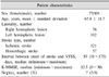
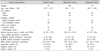
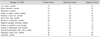
One hundred and seventy-eight subacute stroke patients were included. Their mean age was 67.4 ± 11.7 years. There were 76 patients with right hemispheric lesion and 102 patients with left. 121 patients had ischemic stroke and 57 patients had hemorrhagic stroke. The median interval from onset of stroke to VFSS was 59 days. The median score of K-MMSE was 12.5. Seven patients had hemispatial neglect (Table 1). Numbers of patient in cortical group was 47 (male 31, female 16; right hemispheric lesion 17, left hemispheric lesion 30), subcortical group was 96 (male 50, female 46; right hemispheric lesion 43, left hemispheric lesion 53) and brain stem group was 35 (male 20, female 15; right hemispheric lesion 16, left hemispheric lesion 19) (Table 2). Numbers of patients showed abnormal findings of VFSS in each group are presented in Table 3.
2) Factors affecting dysphagia depending on swallowing phase
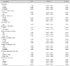
The patients' age, gender, types of stroke, lesion location and laterality, BMI, presence of aphasia and hemineglect, scores of K-MBI and K-MMSE were not associated measured function in oral phase (lip closure, bolus formation, mastication, tongue to palate contact, premature bolus loss, oral transit time of bolus). From the results of VFSS, reduced laryngeal elevation in pharyngeal phase was significantly related to the brain stem lesion (p = 0.045). Pyriform sinus residue after swallowing was associated with aphasia and K-MBI score (p = 0.018). Prolonged pharyngeal delay time was related to brain stem lesion (p = 0.023), ischemic stroke (p = 0.027) and the age of the patient (p = 0.011) (Table 4).
In this study, any pattern of dysphagia was not associated with factors such as patients' gender, lesion laterality, BMI, hemineglect, K-MMSE score.
Discussion
Dysphagia usually occurs when the brain stem or bilateral cortices are injured.17 It is also observed in the patients with unilateral cerebral hemispheric lesion,18 however, the mechanism remained still illusive. According to a previous study, 37% of the patients with unilateral cerebral hemispheric lesion had dysphagia.19 It has been known that there is no correlation between dysphagia and lesion laterality,20 which was in line with our results.
It has been argued that the location of the lesion is more important than the size or laterality of the lesion in predicting aspiration.21 However, most of previous studies showed a contradictory results regarding lesion location. Falsetti et al.22 reported that there was a high incidence of dysphagia in the cortical lesion of non-dominant hemisphere, but neither the subcortical lesion nor the brain stem lesion. Moreover, some other studies demonstrated that brain stem and cerebellar lesions had no statistical significant association with a high incidence of dysphagia.22,23,24 The evidence seems unclear so far, however, if one considers the fact that patients who had brain stem lesions showed abnormal findings not only in the oral phase especially prominent in pharyngeal phase including laryngeal elevation, cricopharyngeal achalasia and prolonged pharyngeal transit time5 as observed in this study, lesion location would be one important factor for risk of dysphagia. Therefore, our results suggests that the brain stem lesion plays a positively associated roles with dysphagia, especially in the pharyngeal phase, even though in the case of controlling the other confounding variables such as the patient's age, gender, associated disease and cognitive dysfunction. None of the other dysphagia variables could be predicted by brain lesion location.
Robbins et al.8 argued that aphasia and hemineglect may also be related to dysphagia along with the lesion location. In addition, according to Schroeder et al.,25 hemispatial neglect was related to initial dysphagia, but aphasia was unrelated to initial dysphagia. However, this study found that in the aphasia patients, the increase of residues of pyriform sinus after swallowing and the risk of dysphagia in the pharyngeal phase was increased by about 3 times, while that hemineglect did not have any relation to dysphagia in both oral phase and pharyngeal phase. According to our result, one need to pay special attention to the patients with aphasia when assessing swallowing difficulty. However, in case of hemineglect, as the number included in this study was too small (7 patients, 4%), it need to be verified further.
It is commonly accepted that aging is one of the important factor leading dysphagia in elderly patients. In older patients, delays in the transfer of the bolus through the oropharynx, and problems in the opening of the upper esophageal sphincter have been reported to occur.26 In our study, age itself was an independent variable in causing dysphagia in the pharyngeal phase.
In this study, K-MMSE score was not associated with any pattern of dysphagia. As it is a screening test for evaluation of cognitive function, it might have not sufficient sensitivity.
There are several limitations in this study. Firstly, the physicians who carried out VFSS were different time to time. Secondly the study design that we used in this study was a retrospective method, which would have problems of accuracy and completeness. Further prospective study in larger cohort of patients would be needed to find out the factors that affect the patterns of dysphagia.
Conclusions
In this study, particularly brain stem lesions showed patterns associated with pharyngeal dysphagia such as reduced laryngeal elevation and prolonged pharyngeal delay time. However, other brain lesions were not associated with any pattern of dysphagia, and no factors were associated with oral dysphagia.
In patients with aphasia and whose independent performance of daily living activities is severely compromised, and in older age groups and ischemic stroke patients, the incidence of pharyngeal dysphagia, such as large amount of pyriform sinus residue, prolonged pharyngeal delay time was increased.
 XML Download