

 PDF
PDF ePub
ePub Citation
 Print
Print


INTRODUCTION
Prosopometamorphopsia is an unusual disorder of face perception in which faces appear distorted to the perceiver, making patients very uncomfortable because features of face look drooped, afloat, protruded, or contracted.1 Usually in prosopometamorphopsia, the whole face looks distorted. However, sometimes distortion is found in some parts of face or one side of face.234567891011 Responsible lesion of unilateral prosopometamorphopsia is usually a lesion of the right hemisphere with a variety of locations of lesion including temporal lobe, occipital lobe, and parietal lobe.3 However, lesion of the left hemisphere has been also reported.7 Hence, the pathophysiologic mechanism of unilateral prosopometamorphopsia is currently unclear. Cases with unilateral prosopometamorphopsia caused by splenial lesion have been very rarely reported.5891011 No such case has been reported in Korean academic journals. Here I report a patient who showed left prosopometamorphopsia after a left splenial infarction recently along with a literature review.
CASE REPORT
A 52-year-old right-handed woman visited our hospital complaining that the left half of people's faces suddenly looked distorted to her a month ago. She complained that the left face of the person (the part on her right side) looked distorted. She stated that the left eye of people looked smaller and inclined compared to their right eye. In addition, the left lip and cheek around it looked swollen to her. When she looked at herself in the mirror, her face in the right visual field looked distorted too. She particularly complained that she could not put on makeup of her lips. She did not complain of amblyopia or visual field defect. During the beginning week in which symptoms occurred, she was hospitalized in another hospital and underwent preservative therapy after taking brain imaging studies. However, there was no significant change in symptoms. She had no particular remarks in medical history or family history. There was no history of smoking or alcohol ingestion. She did not take any drugs prior to the occurrence of the disease. In addition, as a housewife who graduated from high school, she did not have any history of exposing to heavy metals or toxic substances.
On physical examination conducted after hospitalization, vital signs showed that blood pressure of 139/86 mmHg, pulse rate of 82/min, respiration rate of 20/min, and body temperature of 36.0℃. On neurological examination, her consciousness was clear and orientations to time, place, and person were normal. Her visual acuity was normal. Visual field defect was not observed in confrontation test. When watching faces of people in real life or pictures, left prosopometamorphopsia occurred. However, human body or objects or landscape could be normally seen. Some picture similar to prosopometamorphopsia seen in the patient's eyes was chosen by the patient and presented in Fig. 1A. Prosopagnosia was not observed. Callosal disconnection signs including left hand apraxia, left hand anomia, left tactile anomia, left hand agraphia, right hand constructional apraxia, and alien hand were also not observed. Muscular strength of limbs, tone of muscle, sensitive facilities of limbs were normal. Deep tendon reflexes were normal without pathologic reflexes.
Complete blood counts, blood chemistry tests, urinalysis, blood coagulation tests, and serum tumor marker tests were all normal. Her erythrocyte sedimentation rate and C-reactive protein level were also normal. Thyroid function tests, vitamin B12, and folic acid level were in normal range. No abnormal finding was reported in chest X-ray, electrocardiography, or echocardiography. In serum tests, hepatitis B and C were negative. Test for syphilis was also negative. Antinuclear antibody, rheumatoid factor, anti-ds-DNA anti-body, anti-neutrophil cytoplasmic antibody, and other autoimmune antibodies were all normal. Paraneoplastic antibody tests were also negative. Cerebrospinal fluid examination was normal. Oligoclonal band was negative. Her IgG index was 0.47. Visual evoked potential test and electroencephalography were all normal. Automated perimetry did not show visual field defect.
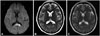
In brain magnetic resonance imaging conducted on the next day of the symptom occurrence, a high signal intensity lesion was observed in the left splenium of the corpus callosum on diffusion-weighted image (Fig. 2A). Apparent diffusion coefficient map was not included in the image data taken at that time. However, intracranial and carotid magnetic resonance angiography was found to be normal. Brain MRI obtained 1 month after onset of the symptom revealed a high signal intensity lesion in the splenium of the corpus callosum on T2-weighted image (Fig. 2B). T1-weighted images showed low signal intensity without contrast enhancement. Gradient echo imaging was normal. There was no abnormal finding in other parts of brain. 99mTc-hexamethyl propylene amine oxime single photon emission computed tomography was performed on Day 5 after onset, which showed normal findings.
Although specific etiology was not found, the patient was diagnosed with unilateral prosopometamorphopsia caused by acute infarction in splenial corpus callosum. Clopidogrel at 75 mg/day was administered and clinical progress was observed in outpatient treatment. The patient's unilateral prosopometamorphopsia was gradually improved. After 3 months, the left eyes of people looked normal to her, but the left lips and cheek around it still looked swollen. After about a year, follow-up brain MRI showed a tissue loss and an atrophic change of the left splenial lesion. No other parenchymal abnormality was observed (Fig. 2C). The extent of unilateral prosopometamorphopsia of the patient was reduced. However, she stated that the left lips and left cheeks of people still looked swollen. The shape of face of other people that the patient drew herself is described in Fig. 1B.
DISCUSSION
Metamorphopsia is the condition in which a patient is aware of objects, but the form, size, color, and depth look different from the real ones. Micropsia (in which objects look smaller than reality) and macropsia (in which objects look bigger than reality) are relatively common forms of metamorphopsia. Prosopometamorphopsia is one of such metamorphopsia. Its causal diseases include cerebral infarction, cerebral hemorrhage, cerebral arteriovenous malformation, brain tumor, seizure, migraine, infectious mononucleosis, and lysergic acid diethylamide or mescaline poisoning in addition to eye disease.19
According to the classical model, neural circuit related to facial perception is mostly included in the ventral occipitotemporal cortex, which is mainly controlled by parts called core system. In the core system, the occipital face area (OFA) in the inferior occipital gyrus, the fusiform face area (FFA) in the middle lateral fusiform gyrus, and face-selective regions in the posterior superior temporal sulcus (pSTS) are included. Although precise functions of these areas are uncertain, some studies have proposed that the OFA is involved in early processing of facial features; the FFA in processing invariant aspects of faces such as facial identity; and the pSTS in the processing of changeable aspects of faces, such as those involved in producing facial expression. In addition to this core system, the areas of intraparietal sulcus, auditory cortex, amygdala, insula, and anterior temporal included in extended system are mobilized to analyze the meaning of information obtained from face, participating in perception process of face. Such face perception processings are known to be particularly dominant in the right hemisphere.1213 Studies using diffusion tensor imaging have demonstrated strong connectivity in white matters between OFA and FFA as well and a significant right-hemisphere predominance. These studies also have demonstrated that splenial fibers connected posterior cortical areas, including striate, extrastriate, and posterior parietal areas and there was greater splenial connectivity from the right hemisphere, particularly to extrastriate cortices.1415
The exact mechanism of prosopometamorphopsia remains unclear. Based on previous studies, face-responsive regions in occipital areas of each hemisphere are involved in building up internal representation of the contralateral side of face. Output pathway from this stage would then be directed towards the right FFA where the integration of information from two sides of face takes place. If there is a disruption in some parts of this pathway, the whole face or one side of the face might have prosopometamorphopsia.347 In the present case, left prosopometamorphopsia might have occurred because the information of the left face of person which is transferred to the right FFA through the splenium after being processed in the face perception areas of the left occipital lobe was interrupted by infarction.
Previous case reports (including present case) of unilateral prosopometamorphopsia caused by splenial corpus callosum lesion are summarized in Table 1. The common characteristics of these cases are metamorphopsia that did not occur in the body or objects in addition to prosopometamorphopsia. Most of them had left prosopometamorphopsia in the right hemifield of patients regardless of the lateralization of splenial lesion except cases reported by Ebata et al.5 and Saito et al.10 The patient reported by Ebata et al. had a right prosopometamorphopsia due to a hemorrhagic lesion which involved the right retrosplenial cortex and might have damaged the pathway from face perception areas in the right occipital lobe to the right FFA. The patient reported by Saito et al. was a left-handed and had a right prosopometamorphopsia because the left-handed patient might have had left hemisphere dominance in face perception areas. It has been reported that most face-sensitive areas identified were right-lateralized in left-handers but FFA was slightly left lateralized in the population of left-handers.16
Although the pathophysiologic mechanism of prosopometamorphopsia is currently unclear, it is thought that unilateral prosopometamorphopsia occurring in the right hemifield of right-handed person may be considered as a callosal disconnection sign caused by a right hemispheric predominance in face perception areas. Such dominant hemisphere-specific disconnection sign is characterized by the fact that neurologic abnormalities are observed in the ipsilateral side of the dominant hemisphere.17 Further studies are needed to investigate the pathophysiologic mechanisms to understand how splenial lesion could cause unilateral prosopometamorphopsia.


 XML Download