

 PDF
PDF ePub
ePub Citation
 Print
Print


Muscle can be protruded through a defect of the fascia into the subcutaneous fat and present clinically as a soft-tissue mass. Although most cases of muscle hernia are asymptomatic and don't require operative treatment, a few patients, often athletes, present with severe pain or cramps require surgery. We report a case of tibialis anterior muscle hernia treated with a local periosteal rotational flap.
CASE REPORT
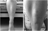
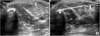
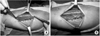
A 19-year-old female admitted to our hospital due to acute subdural hemorrhage after a motorcycle accident. Four months after the accident, she visited our center and complained of palpable mass on her left lower leg, without any external wound or scar. Other hospitals had unsuccessfully attempted to aspirate the mass under the diagnosis of a cystic tumor. However, the mass continued to grow, she had more discomfort and pain sensation over the mass. Especially while she was trying to wear a pant, the mass was prominent and squatting position exacerbated the discomfort (Fig. 1). Dynamic ultrasonography was conducted and a 2.4 cm transverse fascial defect was detected (Fig. 2), showing a herniated tibialis anterior muscle through the defect, especially with dorsiflexion motion of the ankle joint. We diagnosed her with tibialis anterior muscle hernia.
At the first time, support hose was applied and we ordered restrictions and limitations regarding exercise for 1 month, but the patient wanted a surgical treatment as the herniated muscle had not reduce in size.
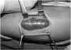
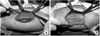
We made a 10 cm longitudinal incision on the anterior aspect of the mass. The tibialis anterior muscle was immediately exposed without a fascial cover of cm ovoid shape, and a pseudomembranous fibrous cover was found on the exposed muscle (Fig. 3). The long duration of these conditions created the retracted fascia rigid state and primary repair was not possible. The anteromedial surface of tibia was exposed by dissection, and the width of the anteromedial side of the tibia was measured as 5 cm, which was sufficient to cover the fascial defect. The length of the periosteal flap was determined to be 1 cm longer than the length of the fascial defect. After sharp incision on the periosteum, meticulous subperiosteal dissection was performed with precautions not to tear the periosteal flap. The flap size was cm and we inverted the periosteal flap and sutured from the tibial side over the defected fascial side (Fig. 4). Fascial defects were covered by the periosteal flap without excessive tension, and the remaining periosteal portion was overlapped and sutured for augmentation (Fig. 5). At 3 weeks after the procedure, the patient could walk on crutches, and after one year, there was no remaining muscle hernia or complications, including pain or cosmetic problems. The patient was satisfied with the result.
DISCUSSION
Muscle hernias are not rare but have received little attention in the medical literature8). A muscle hernia often occurs in the leg and the most common hernia site is the anterior compartment of lower leg1). Physical examination, ultrasonography, and magnetic resonance imaging are used to diagnose this disorder. The efficacy of ultrasonography as a diagnostic tool for muscle hernia has been reported. Dynamic ultrasonography is a non-invasive, highly accurate, and readily available imaging technique for suspected muscle hernia1). Patients with muscle hernia seek medical advice because of painful symptoms, the possibility of a tumor or cosmetic concerns. Asymptomatic hernias require no treatment, and cosmetic problem usually is not an indication for surgery. For patients with moderate to severe symptoms or those in whom conservative treatment has failed to alleviate symptoms, surgical methods can be considered8).
There are several surgical methods of treatment for muscle hernia, including direct repair, fasciotomy8), fascial patch grafting using autologous fascia lata, fascial splitting2) or synthetic mesh6,9). Direct repair is possible when the defect is small and the laxity of the borders permits approximation. But if attempted by force can make repair fail, causing recurrence of the hernia or complication of compartment syndrome2). Some doctors even insisted that a small anterior tibial compartment hernia need not be closed by direct approximation of fascial edge7,10). Some authors suggest fasciotomy as a safe method of surgical treatment1,7). But the fasciotomy is indicated only for small fascial defects, and often results in adhesions between the muscle and the cutaneous scar, causing an evident skin depression upon muscle contracture9). Also it could weaken the muscle fascia and increase the possibility of muscle hernia6). The fascial patch grafting using autologous fascia lata has the weakness of donor site morbidity, causing cosmetic problems9). A reinforcing patch of autologous or synthetic material is another options9). This procedure is simple, and can be used for large defects. But the use of a synthetic patch has drawbacks including cost and risk of graft intolerance to normal surrounding tissue.
The periosteum consists of multipotent mesodermal cells that are capable of differentiating into various types of connective tissue and bone. Histologic examination of the periosteum showed the presence of an outer 'fibrous layer' and an inner'cambium layer'. Because of osteogenic and chondrogenic property, perisoteal flap was studied scientifically and used in orthopaedic surgery actually including ligament reconstruction3). The procedure using auto periosteum was previously introduced4). In 2009, Marić et al5) already reported thress cases treated with similar method. But to the author's knowledge, this is the first description of local periosteal rotational flap procedure concerning this problem written in English. Considering the medial side of the tibia, Siliprandi et al9), in 1993, stated that the periosteum could not be used to cover a large defect of more than 3 to 4 cm. Miniaci and Rorabeck8), in 1987, mentioned the risk of compartment syndrome. However, to date, there are no case reports of compartment syndrome with the local periosteal rotational flap. We think that preventing excessive tension is important and recommend this technique for treating defects those size do not exceed periosteal width. The contraindication, however, does not include the length of a defect. Cases of small sized hernia of the tibialis anterior muscle, including this case, are frequently reported, and therefore this method could be used in many cases4). The local periosteal rotational flap method has several advantages compared to previous techniques. It could be used in relatively large defect. It has no donor site morbidity and skin irritation after treatment. It is simple and can be done with only single incision. It doesn't need additional cost and has no fear of rejection of graft material. If the fascial defect is small enough to be overwrapped by the periosteal flap (Fig. 5B), it may provide more strength to a defective area.
Even though the number of cases in this study was small, the local periosteal rotational flap for tibialis anterior muscle hernia provided satisfactory outcome without serious complications and could be an alternative option to conventional operative treatments.
 XML Download