

 PDF
PDF ePub
ePub Citation
 Print
Print


Abstract
The purpose of this study was to provide basic biometric data on Korean adults through magnetic resonance imaging (MRI)-based measurements of the distances between the apex of sacral hiatus (SH) and the termination of dural sac (DS), and between SH and conus medullaris (CM) because they are critical to the performance of epidural neuroplasty.
A total of 200 patients (88 males and 112 females) with back pain, who had no spine fracture, significant spinal deformity, and spondyloisthesis were selected for this study. The subjects were of mean age 54.3 (20∼84) years and mean height 161.3 cm (135∼187). T2-weighted MRI images were used for correlation analysis to evaluate the relationships between the distances, and variables such as sex and height.
In all patients, the mean distance between SH and DS was 62.8±9.4 mm and the mean distance between SH and CM was 232.2±21.8 mm. The minimum distance and the maximum distance between SH and DS were 34.8 mm and 93.9 mm respectively, and the minimum distance and the maximum distance between SH and CM were 155.0 mm and 284.0 mm respectively.
In female patients, both the distances between the SH and DS, and between SH and CM were shorter when compared to those of the male patients (p<0.05). Both the distances between SH and DS and between SH (p<0.01). The results of this study will provide a useful and CM showed a significant correlation with height biometric data on the distances between SH and DS and between SH and CM in Korean in ensuring clinical safety and in the development of more effective catheterization techniques for epidural neuroplasty in Korean.
References
1. Racz G, Heavner J, Trescot A. Percutaneous lysis of epidural adhesions: evidence for safety and efficacy. Pain Pract. 2008; 8:277–86.
2. Hsu E, Atanelov L, Plunkett A, Chai N, Chen Y, Cohen S. Epidural lysis of adhesions for failed back surgery and spinal stenosis: factors associated with treatment outcome. Anesth Analg. 2014; 118:215–24.
3. Erdine S, Talu GK. Precautions during epidural neuroplasty. Pain Pract. 2002; 2:308–14.

4. Ho KY, Manghnani P. Acute monoplegia after lysis of epidural adhesions: A case Report. Pain Pract. 2008; 8:404–7.
5. Bogduk N. The sacrum. Clinical anatomy of the lumbar spine and sacrum. 4th ed.Philadelphia: Elsevier Churchill Livingstone;2005. p. 59–61.
6. Senoglu N, Senoglu M, Oksuz H, Gumusalan Y, Yuksel KZ, Zencirci B, et al. Landmarks of the sacral hiatus for caudal epidural block: an anatomical study. Br J Anaesth. 2005; 95:692–5.
7. Sekiguchi M, Yabuki S, Satoh K, Kikuchi S. An anatomic study of the sacral hiatus: a basis for successful caudal epidural block. Clin J Pain. 2004; 20:51–4.
8. Demiryürek D, Aydingöz Ü, Akşit M, Yener N, Geyik P. MR imaging determination of the normal level of conus medullaris. Clin Imaging. 2002; 26:375–7.
9. Louis R. Topographic relationship of the vertebral column, spinal-cord, and nerve roots. Anat Clin. 1978; 1:3–12.
10. Kwon SW, Kim TS, Kim HS, Rhyu IJ. The Tip Level of the Conus Medullaris by Magnetic Resonance Imaging and Cadaver Studies in Korean Adults. Korean J Phys Anthropol. 2016; 29:47–51.
11. Macdonald A, Chatrath P, Spector T, Ellis H. Level of termination of the spinal cord and the dural sac: a magnetic resonance study. Clin Anat. 1999; 12:149–52.
12. Becker SH, Arenson RL. Cost benefits of picture archiving and communication system. J Am Med Assoc. 1994; 1:361–71.
13. Deyo RA, Loeser JD, Bigos SJ. Herniated lumbar intervertebral disk. Ann Intern Med. 1990; 112:598–603.
14. Shin SW. Low back pain: Review of anatomy and pathophysiology. J Korean Med Assoc. 2006; 49:656–64.
15. Suk SI. Lumbar disc disease. Spinal surgery. Revised edition. Seoul: Newest Medical Journal;2004. p. 218–20.
16. Modic MT, Pavlicek W, Weinstein MA, Boumphrey F, Ngo F, Hardy R, et al. Magnetic rresonance imaging of intervertebral disk disease: Clinical and pulse sequence considerations. Radiology. 1984; 152:103–11.
17. Barson AJ. The vertebral level of termination of spinal cord during normal and abnormal development. J Anat. 1970; 106:489–97.
18. Kim DC. Anatomical studies on the vertebral column and spinal cord of korean subjects: Part Ⅱ. Study on the adult. Seoul J Med. 1961; 2:149–63.
19. Joo SP, Kim SH, Lee JK, Kim TS, Jung S, Kang SS, et al. The variation of position of the conus medullaris in korean adults: A magnetic resonance imaging study. J Korean Neurosurg Soc. 2001; 30:451–5.
20. Park SJ, Choi WS, Kim EJ, Oh JH, Yoon Y. Normal variation of lumbosacral thecal sac on MR. J Korean Radiol Soc. 1996; 34:565–69.
21. Ko HY, Kim K, Kim HJ. Normal variations of the spinal cord termination. J Korean Acad Rehabil Med. 1998; 22:1040–43.
22. Ko HY, Park JH, Kim HJ, Kim K. Variations of end level of the dural sac. J Korean Acad Rehabil Med. 1999; 23:805–8.
23. Nasr AY. Vertebral level and measurements of conus medullaris and dural sac termination with special reference to the apex of the sacral hiatus: anatomical and MRI radiologic study. Folia Morphol. 2016; 75:287–99. doi:. DOI: 10.5603/FM.a2016.0004.
24. Mustafa MS, Mahmoud OM, El Raouf HH, Atef HM. Morphometric study of sacral hiatus in adult human Egyptian sacra: Their significance in caudal epidural anesthesia. Saudi J Anaesth. 2012; 6:350–7.
Fig. 1.
Determination of the exact measurement of position in relation to the location and the type of variation of the sacral hiatus (SH): (A) Identification of the position of SH using bone setting sagittal images with CT, and (B) Determination of the exact location of measurement by using T2-weighted MR images (arrows; Apex of the SH).
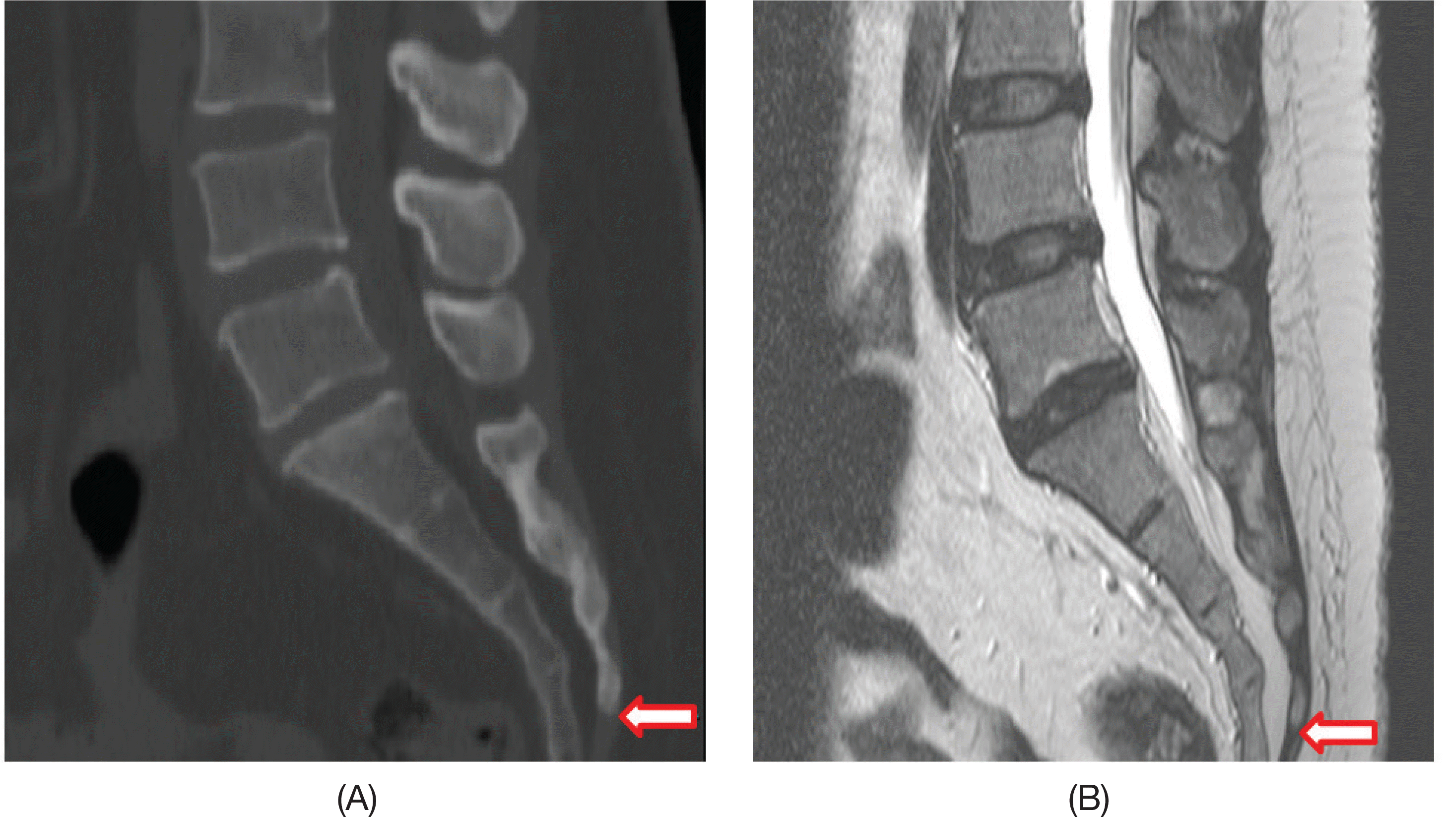
Fig. 2.
Schematic illustration of the measurement methods: (A) Identification of the position of the sacral hiatus the distance between SH and the termination of dural sac (SH). (B) Measurement of (DS). (C) Measurement of the distance between SH and the conus medullaris (CM).
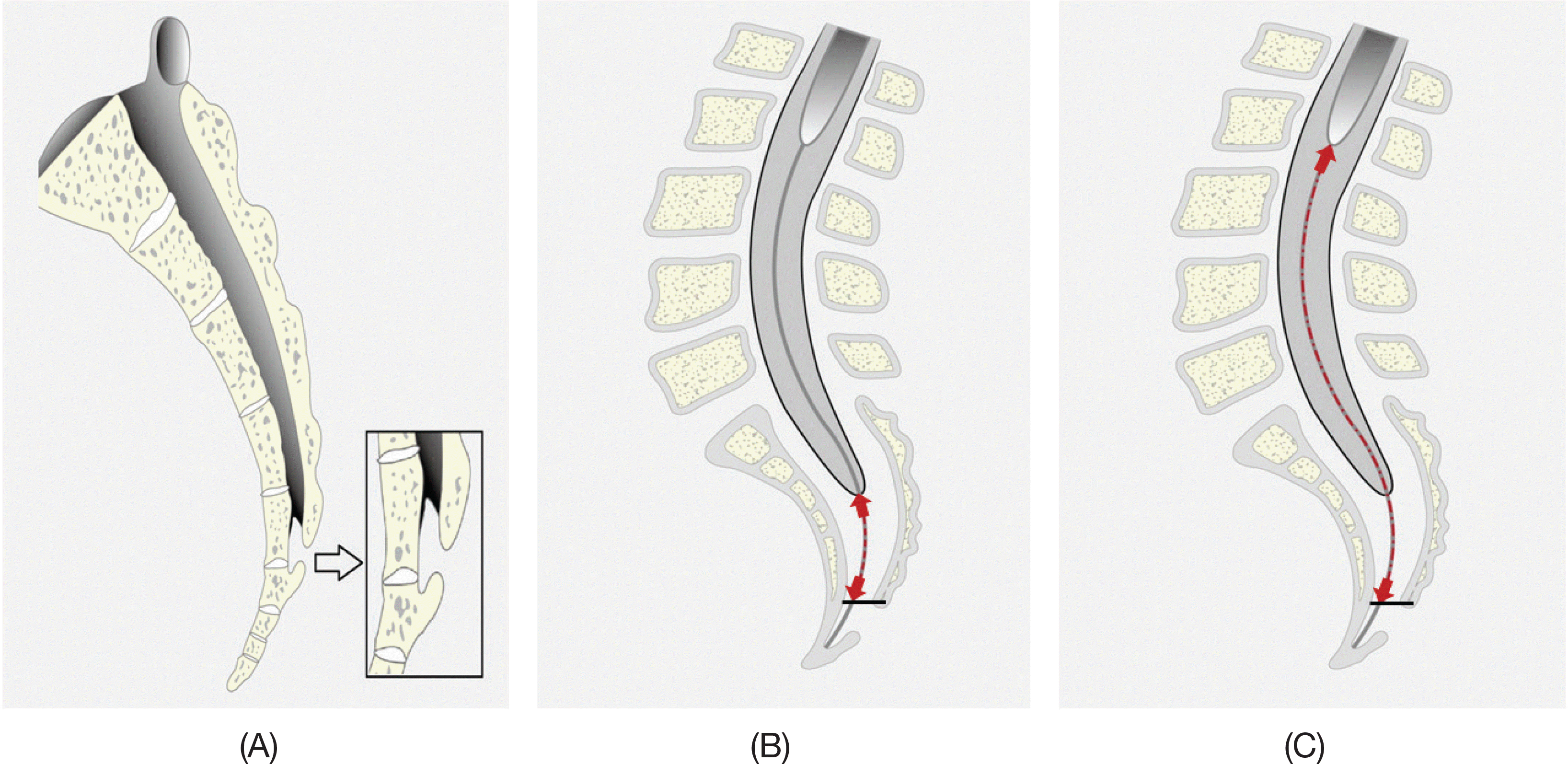
Fig. 3.
MRI T2-weighted, midsagittal plane images using PACS (picture archiving communication system) were measured along the vertebral canal: (A) The position of the sacral hiatus (SH), termination of dural sac (DS) and conus medullaris (CM). (B) The distance between SH and DS. (C) The distance between SH and CM.
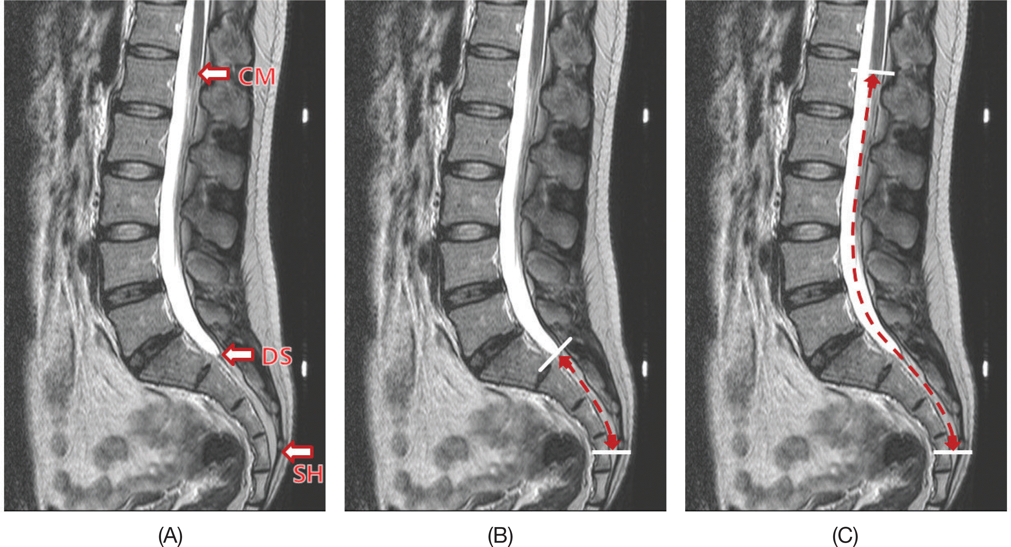
Fig. 4.
Location of the Apex of the sacral hiatus (SH). The incidences of the apex of SH located at the level of S3 to S5 vertebrae were 9.5% at the level of S3, 63.5% at the level of S4, and 27% at the level of S5.
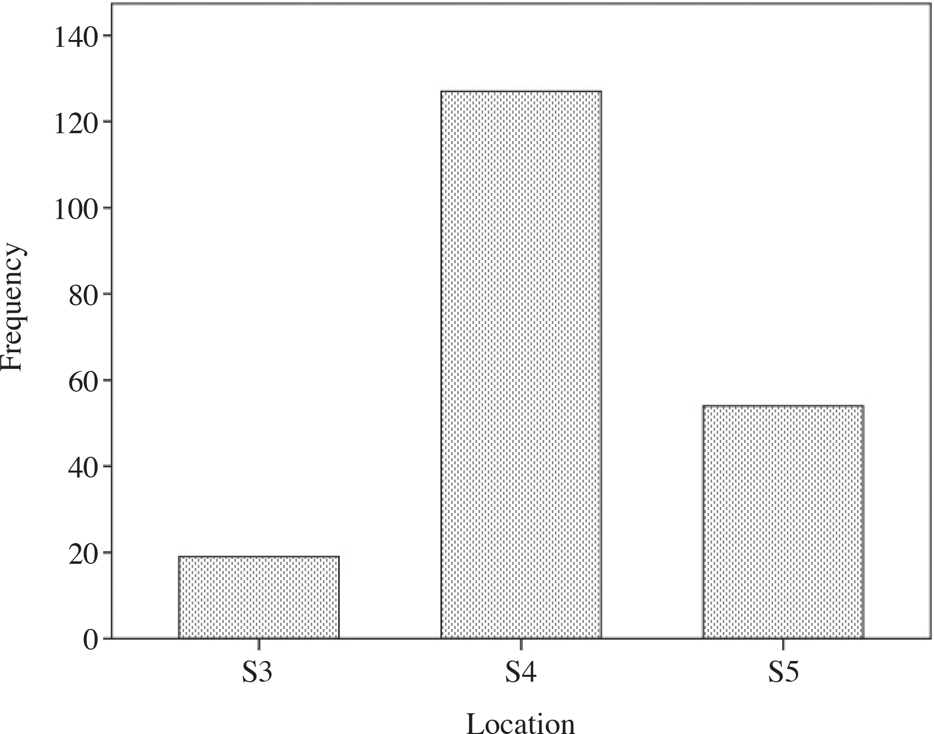
Fig. 5.
Histogram showing the frequency of the vertebral level of the apexes of the sacral hiatus in male and female subjects. 5.5% of the male subjects, and 4% of the female subjects were located at the level of S3. 28% of the male subjects, and 35.5% of the female subjects were located at the level of S4. 10.5% of the male subjects, and 16.5% of the female subjects were located at the level of S5.
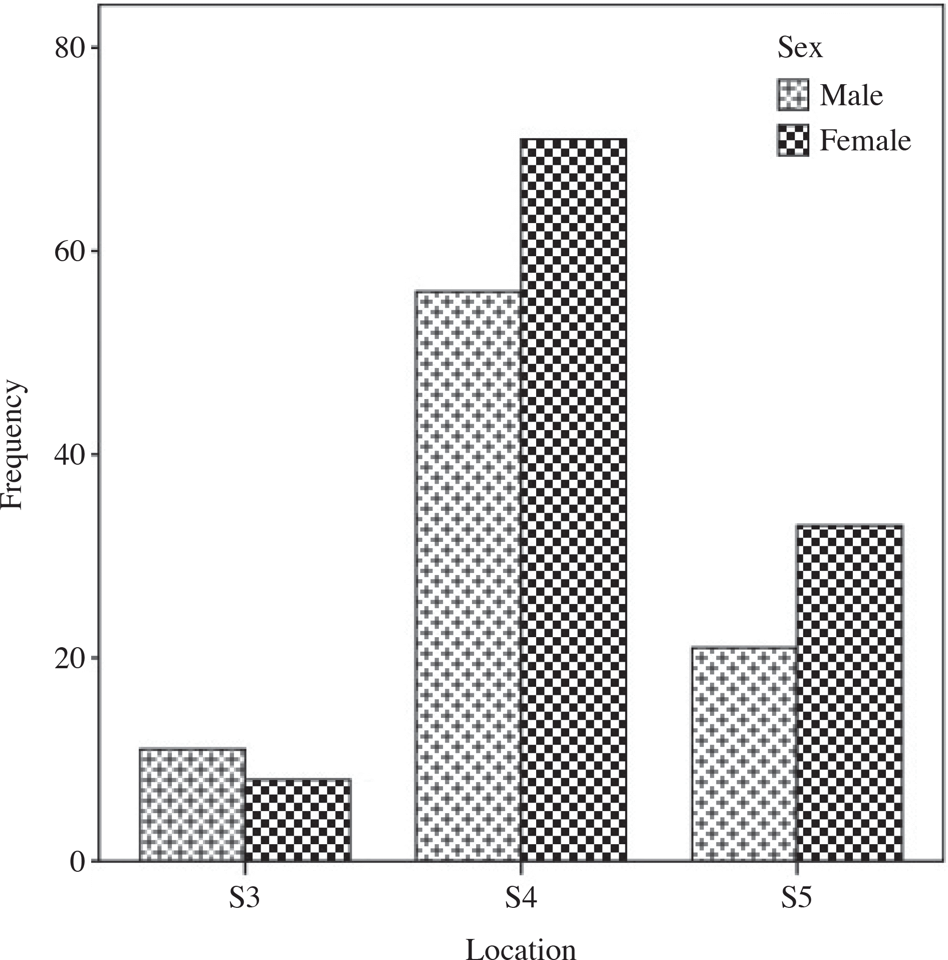
Fig. 6.
Histogram of the frequency of the vertebral level of the apexes of the sacral hiatus among the different height groups in (≤149 both male and female subjects. 1.5% of the height group (150∼159 cm), 3% of the height cm), 2% of the height group (160∼169 cm), and 3% of the height group (≥170 cm) were group (≤149 cm), located at the level of S3. 11.5% of the height group (150∼159 cm), 18% of the height group 22.5% of the height group (160∼169 cm), and 11.5% of the height group (≥170 cm) were (≤149 cm), located at the level of S4. 3.5% of the height group (150∼159 cm), 13% of the height group 6% of the height group (160∼169 cm), and 4.5% of the height group (≥170 cm) were located at the level of S5.
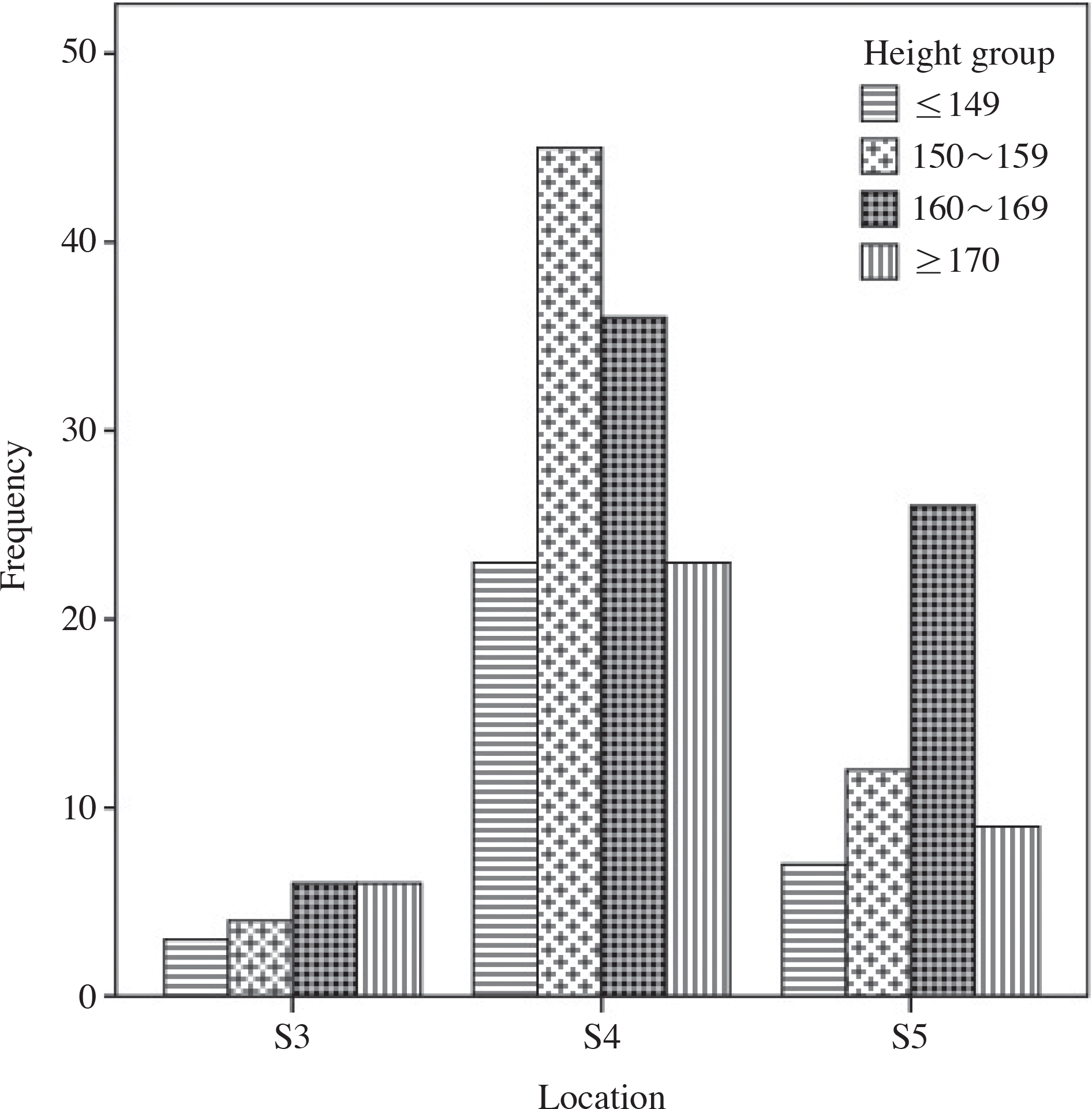
Fig. 7.
Histogram of the distances between the sacral hiatus (SH) and termination of dural sac (DS), and between SH and conus medullaris (CM) according to the sex. (A) The distance between SH and DS. (B) The distance between SH and CM. The data were analyzed by t-test and were considered significant at p<0.05.

Fig. 8.
Vertical box plots of the distances between the sacral hiatus (SH) and termination of dural sac (DS), and between SH and conus medullaris (CM) according to the height and sex. (A) The distance between SH and DS. (B) The distance between SH and CM. The data were analyzed by one way ANOVA.
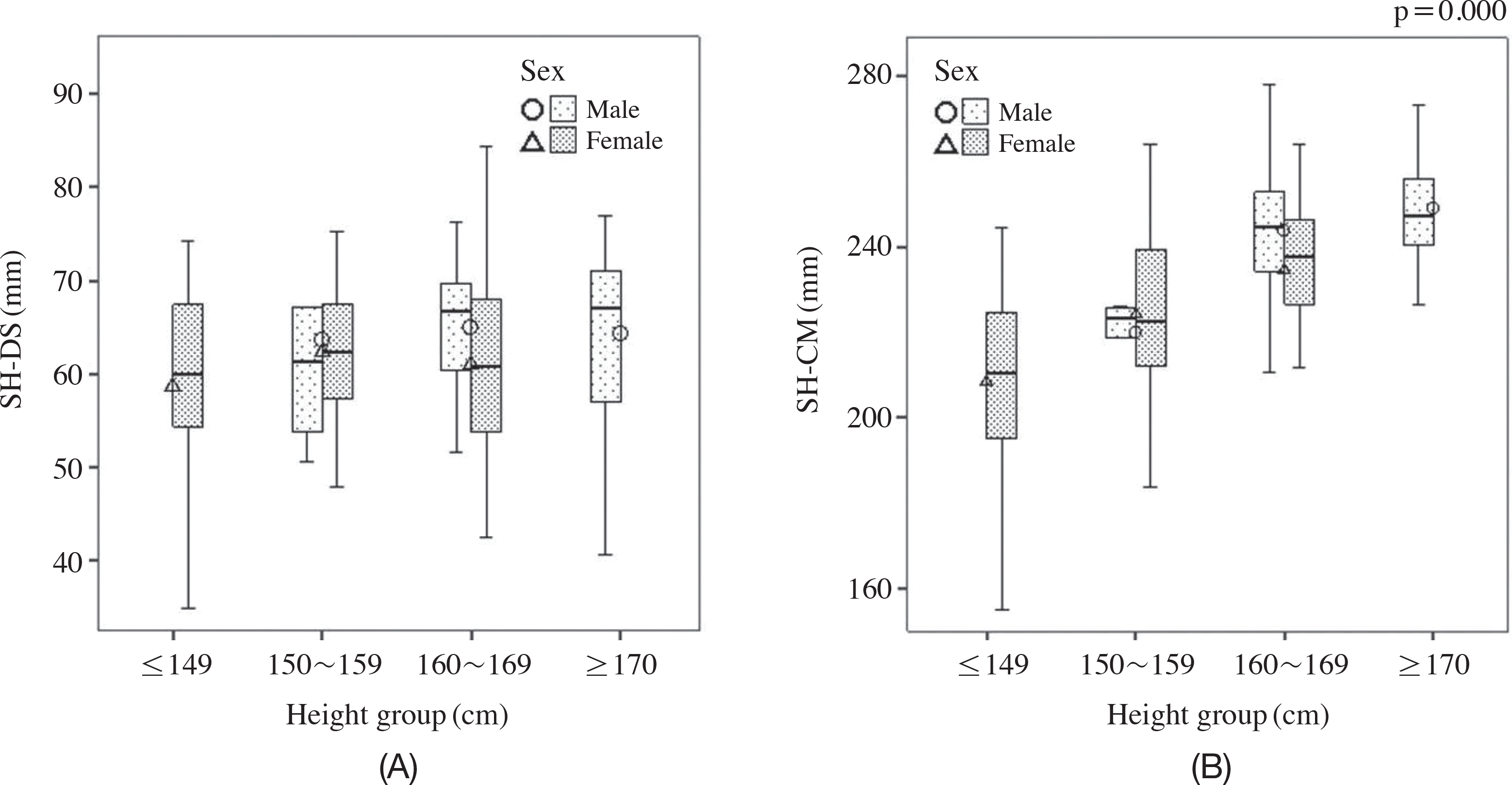
Fig. 9.
Vertical box plots of the distances between the sacral hiatus (SH) and termination of dural sac (DS), and between SH and conus medullaris (CM) according to the height. (A) The distance between SH and DS. (B) The distance between SH and CM. The data were analyzed by correlation coefficient and were considered significant at p < 0.01.
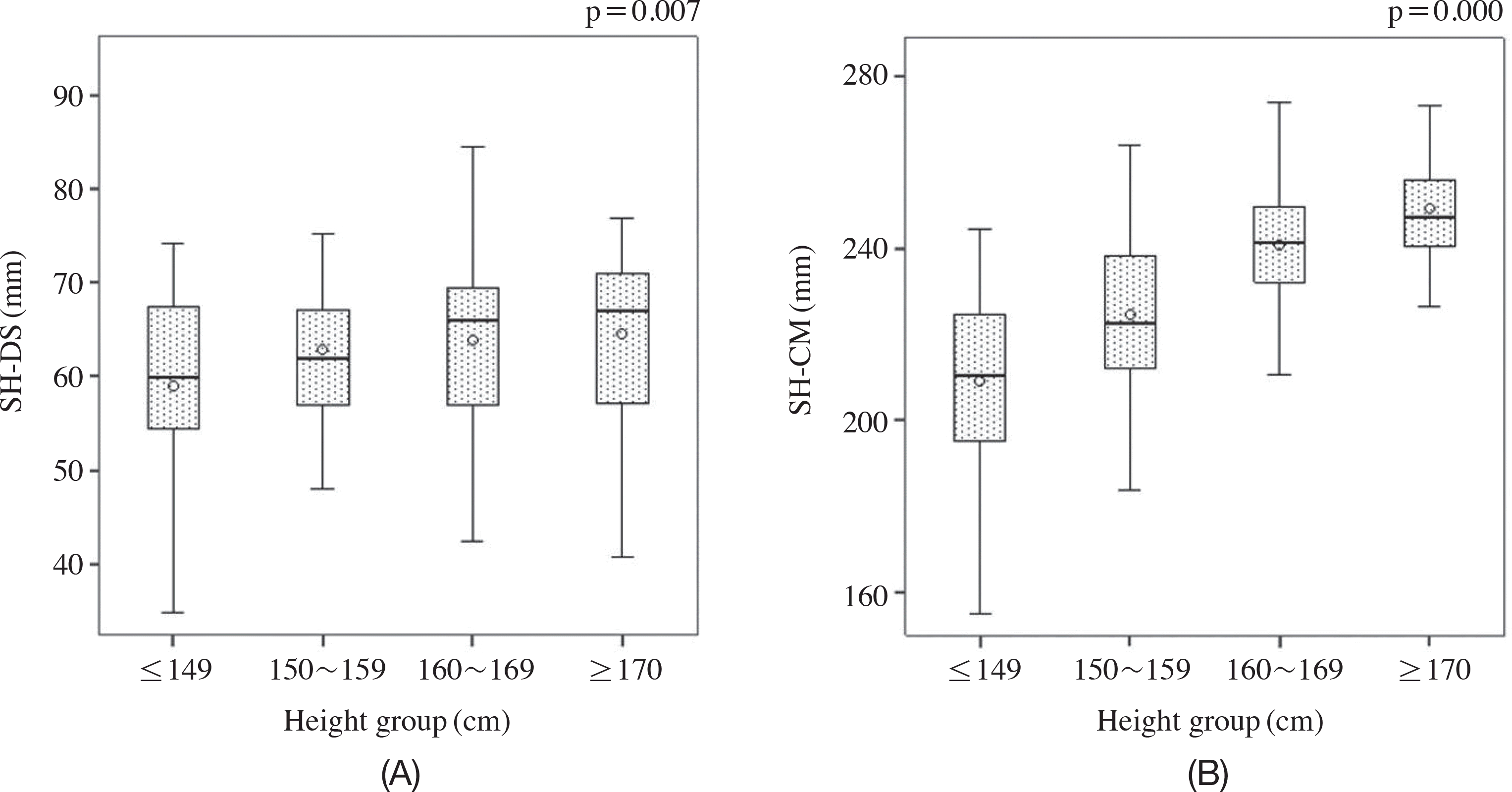
Table 1.
Basic information about research subjects
Table 2.
Comparison of the distance between SH and DS with that between SH and CM according to sex
| Sex | N | Mean±SD (mm) | t | p-value | Total mean±SD (mm) | |
|---|---|---|---|---|---|---|
| SH-DS∗ |
Male Female |
88 112 |
64.7±8.8 61.3±9.6 |
2.612 | 0.010 | 62.8±9.4 |
| SH-CM† |
Male Female |
88 112 |
244.5±16.0 222.5±21.0 |
8.159 | 0.000 | 232.2±21.8 |
Table 3.
Comparison of the distance between SH and DS with that between SH and CM according to height
Table 4.
Analysis on Pearson's correlation coefficients of heigh and location for the distances from SH to DS and CM
| N | Pearson correlation | p-value | ||
|---|---|---|---|---|
| SH-DS | Height | 200 | 0.189∗ | 0.007 |
| Location† | 0.808 | 0.260 | ||
| SH-CM | Height | 0.636∗ | 0.000 | |
| Location† | –0.037 | 0.602 |
Table 5.
The frequency of different locations of the base of the sacral hiatus in relation to sacral vertebrae
 XML Download