

 PDF
PDF ePub
ePub Citation
 Print
Print


Yu Ryang Jang1 , Myeung Ju Kim2
, Myeung Ju Kim2
Abstract
The Ministry of National Defense of Agency for KIA Recovery and Identification (MAKRI) launched the Korean War casualty excavation project to retrieve war remains at the Arrowhead Ridge in the DMZ by affecting the mood of peace-building and inter-Korean tension-reducing, and uncovered possible Korean War casualty's remains. The present case of excavated bones was well-preserved and the rate of preservation was more than 97%. As an identification results of this case, the ethnicity, age, stature was estimated as European, 16–19 years old, 163–169.5 cm respectively. Schmorl's nodes and partial lumbarization of the sacrum were observed in the anthropological and forensic examination. Multiple Schmorl's nodes as various sizes were localized in the upper and lower surface of the vertebral endplate from lower thoracic vertebrae (T8–T12) to lumbar vertebrae (L1–L5). A partial lumbarization of S1 was also observed in the sacrum. In this case, we suggested the hypothesis that Schmorl's node and partial lumbarization of S1 can simultaneously occur even in the young age as a course of functional adaptation in regarding to the morphological features of the constituent elements of the vertebrae. Further studies for the biomechanical mechanism of Schmorl's node and lumbarization of S1 in various ethnic groups of large population will reveal more about the relationship between the morphological features of the vertebrae and the bony lesions.
Figures and Tables
Fig. 1
The Korean War casualty remains of present case excavated from the Arrowhead Ridge in DMZ. A. The uncovered and exposed human skeleton on the spot. B. Well preserved human skeleton and measure scale.
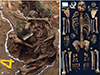
Fig. 2
Lower thoracic vertebrae (T8–T12) and Lumbar Vertebrae (L1–L3) were sequentially arranged from left to right direction. Upper and lower row showed the superior and inferior view of each vertebra, respectively. Various sizes of Schmorl's nodes were found in the upper and lower surface of vertebral endplate in each vertebra, notably L2, L3. The round shape of vertebral body was also well recognized in the thoracic vertebrae.

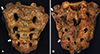
Fig. 3
Sacrum showing partial lumbarization of first sacral segment (S1). A. ventral surface of sacrum. B. dorsal surface of sacrum. The presence of distinct separation was notably observed between the S1 and S1 body. Especially, the lower margin of auricular surface was situated at a lower level (S3) than normal site.
References
1. Yang YJ, Nam JO. Korean War History. Ridge battle and Armistice Agreement. Vol. 3. Ministry of National Defense Military Compilation Laboratory;2015. p. 82–87.
2. Abbas J, Hamoud K, Peled N, Hershkovitz I. Lumbar Schmorl's Nodes and Their Correlation with Spine Configuration and Degeneration. Biomed Res Int. 2018; 2018:1574020.

3. Cho HC, Bae GD, Lee YC, Kim KS. Prevalence of Lumbosacral Transitional Vertebrae in Korean. J Korean Radiol Soc. 1990; 26:1244–1248.
4. Plomp KA, Roberts CA, Viðarsdóttir US. Vertebral morphology influences the development of Schmorl's nodes in the lower thoracic vertebrae. Am J Phys Anthropol. 2012; 149:572–582.
5. Cheng JS, Song JK. Anatomy of the sacrum. Neurosurg Focus. 2003; 15:E3.
6. Rowbotham SK, Blau S, Hislop-Jambrich J. Recording skeletal completeness: A standardised approach. Forensic Sci Int. 2017; 275:117–123.
7. Lovejoy CO, Meindl RS, Pryzbeck TR, Mensforth RP. Chronological metamorphosis of the auricular surface of the ilium: a new method for the determination of adult skeletal age at death. Am J Phys Anthropol. 1985; 68:15–28.
8. Buikstra JE, Ubelaker DH. Standards for data collection from human skeletal remains. Arkansas Archeological Survey Research Series No. 44. Fayetteville: 1994. p. 16–19.
9. Vigilant L, Stoneking M, Harpending H, Hawkes K, Wilson AC. African populations and the evolution of human mitochondrial DNA. Science. 1991; 253:1503–1507.
10. Malaspina P, Persichetti F, Novelletto A, Iodice C, Terrenato L, Wolfe J, et al. The human Y chromosome shows a low level of DNA polymorphism. Ann Hum Genet. 1990; 54:297–305.
11. Raxter MH, Auerbach BM, Ruff CB. Revision of the Fully technique for estimating statures. Am J Phys Anthropol. 2006; 130:374–384.
12. Masharawi Y, Salame K, Mirovsky Y, Peleg S, Dar G, Steinberg N, et al. Vertebral body shape variation in the thoracic and lumbar spine: characterization of its asymmetry and wedging. Clin Anat. 2008; 21:46–54.
13. Mahato NK. Morphological traits in sacra associated with complete and partial lumbarization of first sacral segment. Spine J. 2010; 10:910–915.
 XML Download