

 PDF
PDF ePub
ePub Citation
 Print
Print


Introduction
The interaction between orthodontic forces and the periodontal ligament leads to an inflammatory phenomenon that induces apical resorption by clastic activity1 without clinical symptoms. This phenomenon is known as external apical root resorption (EARR), and it is an undesirable2 and irreversible side effect of orthodontic treatment.
The reported occurrence of EARR is between 48% and 66% according to radiographic studies and more than 90% based on histologic analyses.34 Fortunately, most cases exhibit resorption of no greater than 1 mm, which does not impair tooth function. However, higher degrees of root shortening are observed in approximately 8% of patients one year after orthodontic treatment.5
Several factors are associated with EARR, including increased treatment duration, direction of tooth movement, the loading regimen,2 tooth extraction,1 the type of the tooth and the malocclusion, and patient-related factors including certain systemic conditions, age, and gender.4
The early detection of initial resorptive lesions during orthodontic treatment is essential for identifying teeth at risk of severe resorption.678 The subsequent interruption of active treatment can help to reduce adverse outcomes during later stages of treatment,7 as well as avoiding or limiting the potential damage to the patient.9
The tools most commonly used to detect EARR are periapical18 and panoramic radiographs.10 This is probably due to the frequent use of these exams during the stages of orthodontic treatment1112 and the good cost-benefit outcomes for the patient. Nevertheless, the visual comparison of radiographs before and after orthodontic treatment to measure the EARR is subject to discrepancies in clinical practice, and this technique is not able to detect minor changes in sequential images.1314
Moreover, both radiographic techniques have limitations. Panoramic radiographs show a substantial amount of magnification and do not allow the clear visualization of the premaxilla.11 Although they exhibit much less distortion,11 periapical radiographs require a certain degree of root shortening to have taken place before it is detectable visually on the radiograph.1214
In order to overcome these problems, cone-beam computed tomography has been suggested as an alternative to periapical and panoramic radiographs. The scientific literature indicates that this technique is suitable for the detection of early EARR.112 However, this exam makes orthodontic treatment more expensive and subjects the patient to an additional dose of radiation.
As an alternative, this paper proposes a posteriori registration and subtraction of radiographic images and the use of computerized techniques to quantify the EARR using periapical radiographs. The procedure of a posteriori registration and subtraction corrects the discrepancies of the geometric projection and equalizes the density and the contrast of the radiographic images before and after treatment, enabling the comparison of the two radiographs and improving the sensitivity and the accuracy of the evaluation, which is performed using specific software.
Although this method has been extensively tested in vitro in extracted teeth with simulated resorption,571516171819 the use of periapical radiographs from actual orthodontic treatments,620 as proposed in this paper, is not a common feature of the dental literature on digital subtraction (DSR) radiography and EARR.
The null hypotheses tested were that a posteriori registration and subtraction of periapical radiographic images cannot be used to quantify the EARR following orthodontic treatment and that patient-related factors (gender and age) and treatment-related factors (tooth extraction, use of intermaxillary elastics, and duration of orthodontic treatment) do not affect the amount of root resorption.
Therefore, the objective of this retrospective study was to evaluate whether a posteriori registration and subtraction of radiographic images could be used to quantify apical root resorption in maxillary permanent central incisors after orthodontic treatment, as well as to determine whether the EARR was related to the parameters involved in the treatment.
Materials and Methods
This study was approved by the Ethical Committee of São Paulo State University (Campus of São José dos Campos) under protocol #041/2009.
Sample selection
A careful analysis of 300 orthodontic clinical records from patients treated by dentists who attended a postgraduate dental education program in orthodontics was performed. The inclusion criteria were: orthodontic treatment with the standard edgewise technique; no history of trauma, wear, or endodontic treatment in the maxillary central incisors; complete radiographic exams, including baseline and final periapical radiographs of the maxillary central incisors and cephalometric analysis; the absence of syndromic or skeletal disorders; and complete root formation in the maxillary central incisors. After this screening, 79 patients were selected. The sample number was above the calculated required sample size (n=64; α=0.5; 1- β=0.9), but all the selected patients were kept in the sample to compensate for possible losses during the study.
Radiographic analysis: sequence of procedures
Periapical radiographs were taken using the modified parallel technique and standardized exposure parameters: Heliodent 70 (Siemens, Erlangen, Germany) X-ray unit, 70 kVp, 7 mA, 0.4 seconds. The radiographs were digitalized using a scanner with transparency adapter (HP Scanjet G4050, Hewlett-Packard, Palo Alto, CA, USA) with a resolution of 300 dpi and 8-bit gray scale. The images were stored as maximum-quality TIFF format files.
All the images were imported into the Regeemy Image Registration and Mosaicking 0.2.43-RCB software (DPIINPE, São José dos Campos, São Paulo, Brazil). This software provides image registration and subtraction algorithms (i.e., it corrects geometric discrepancies, equalizes the contrast of two sequential radiographs to make them comparable, and subtracts the analog pixels values from two sequential images).
The radiographic image obtained at baseline (before orthodontic treatment) was termed Image 1 and used as the reference image. The radiographic image obtained after orthodontic treatment was referred to as Image 2.
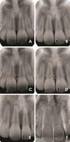
Reference points (fixed points in both radiographs) were selected on both images manually (Fig. 1). These reference points served as coordinates for the software to align and correct the geometry of the second image according to the reference image (Image 1). Clearly distinguishable structures were selected as reference points, such as the cement-enamel junction and the incisal margin of the maxillary central incisors.
This process generated Image 3, which was the retrospectively corrected form of Image 2. The quality of the image correction was visually determined using the image subtraction routine (Image 1-Image 3). The image registration was considered adequate when the structural noise on the teeth of interest was reduced to the lowest possible level, as indicated by a minimal or nonexistent discrepancy in the geometrical position between images, which appears as brighter or darker shades of gray in the subtracted radiograph.
Therefore, the corrected image (Image 3) became the new follow-up radiographic image, since small differences in projection angles during exposure or contrast/density during processing were corrected (Fig. 1).
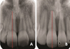
The evaluation of EARR was performed using Image 1 (baseline) and Image 3 (the corrected image) (Fig. 2). The measurement of the long axis of the central maxillary incisors was done using the UTHSCSA Image Tool (University of Texas Health Science Center at San Antonio; http://compdent.uthscsa.edu/dig/itdesc.html). The length of the tooth was considered to be the distance between the root apex and the incisal edge at a specific point corresponding to the mean distance between the mesial and distal angles. The above-described protocol was carried out separately for the right and left maxillary central incisors.
The complete procedures of a posteriori registration and subtraction and DSR were completed by one experienced investigator trained in this methodology. The measurement of the length of the tooth was repeated three times and the final result consisted of the mean of the three evaluations. Intraexaminer agreement was calculated using Cohen's kappa (k=0.87).
The EARR in the central maxillary incisors was determined by the difference between baseline (Image 1) and post-treatment (Image 3) tooth lengths. These values were obtained in pixels. The difference between the values before and after treatment (mean EARR) was obtained and described in terms of pixels and relative root resorption (%).
Evaluation parameters
In order to identify possible correlations with factors that are commonly associated with EARR, data regarding patient characteristics such as gender and age were collected from the clinical records. The features involved in the orthodontic treatment that were considered for analysis were the need for maxillary first bicuspid extraction, the use of elastics, and the treatment duration. The measures 1.NA (angle formed by the maxillary incisor long axis and the nasion line) and 1-NA (linear distance between the most anterior point of the maxillary central incisor and the NA line) were also obtained, since these are part of the cephalometric analysis that determines the dental pattern of the maxillary incisors.
Statistical analysis
The obtained data showed a normal distribution (Shapiro-Wilk). Therefore, the comparisons between mean tooth length, 1.NA, and 1-NA before and after orthodontic treatment were made using the paired t-test. The Student's t-test was used to compare the parameters of gender, age, tooth extraction, elastics use, and treatment duration. The statistical analysis was conducted using SigmaPlot 12.0 (Systat Software Inc, San Jose, CA, USA), with the significance level set at p=5%.
Results
The final sample was composed of 79 patients (mean age, 13.5±2.2 years; range, 10-19 years; 22 males and 57 females). The mean duration of the treatment was 25.8±6.2 months (range, 10-38 months).
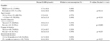
The tooth length before and after orthodontic treatment was 302.9±30.3 pixels and 287.2±32.3 pixels for the right maxillary central incisors; and 302.4±30.2 pixels and 287.2±31.1 pixels for the left maxillary central incisors. No significant differences were observed when the right and left central incisors were compared (p>0.05), and all subsequent analyses were therefore made using the mean of the measurements of the left and right incisors. The mean EARR observed was 15.4±12.1 pixels, representing a resorption of 5.1% (Table 1).
The mean length of the maxillary central incisors, as well as the mean values of the cephalometric measures 1.NA and 1-NA before and after orthodontic treatment are shown on Table 1. These parameters exhibited significant decreases after treatment.
No differences in the mean EARR were observed according to gender, age, the use of elastics, or treatment duration. The only parameter that influenced the patient's mean EARR was the need for tooth extraction as part of orthodontic treatment (Table 2).
Discussion
The data obtained in this study allowed to reject the first hypothesis tested, since a posteriori registration and subtraction of periapical radiographic images performed using specific software was able to quantify the EARR after orthodontic treatment. The second hypothesis was partially rejected, because the parameter of tooth extraction influenced the extent of root resorption.
In our sample, all the patients exhibited EARR to some degree. This finding was expected and has been widely documented in the literature.12469121721222324 The most commonly affected teeth are the maxillary incisors,3252627 which is why this paper evaluated resorption in this specific type of tooth.
Most previous studies investigating EARR used the Malmgren scale to evaluate EARR.9122829 The scale consists of four scores, varying from no resorption to resorption beyond the apical third of the tooth. It is considered a subjective method, and the analysis of two radiographs taken at a temporal interval may introduce some bias, reflecting the instruction, training, and experience of the examiner.513 Additionally, the diagnosis of root resorption by the comparison of periapical radiographs is only possible after five to six months.5
The use of a posteriori registration and subtraction overcame these drawbacks. The accuracy of the DSR method has been confirmed by in vitro studies using extracted tooth.57816 A posteriori registration and subtraction of periapical radiographic images, the accuracy of which was checked with DSR, has been proven to be a method that can quantify small changes associated with EARR in vivo; in our study, it was possible to diagnose early root loss (as reflected by changes as small as 0.6 pixels), and our technique corrected for possible changes in tooth position resulting from orthodontic treatment.26 In daily practice, this process can be performed easily. However, the standardization of the follow-up periapical radiographs should be further developed, since the accuracy of a posteriori registration may have been higher if the vertical and horizontal angulations did not show variations greater than 20° and 10°, respectively.5
Our results showed that the length of the maxillary incisors diminished by a mean of 15.4±12.1 pixels or by approximately 5.1%. These values were measured in pixels and as relative root resorption (%) because these measurement units overcome the inherent variation of the direct measurement of root length,30 and this is probably the best way to standardize the results, allowing the fairest comparisons between different papers that evaluate EARR in a quantitative manner.
Higher mean values have been reported when panoramic radiographs were evaluated (19.5±12.6 pixels),26 as well as periapical radiographs (mean EARR percentage of 9.77% after 12 months of orthodontic treatment).31 This may be expected, since the evaluation of EARR using panoramic radiographs tends to overestimate the amount of tooth loss by 20% or more when compared to periapical radiography,1112 and the evaluation using periapical radiographs without geometric correction may induce shortening or lengthening of the image, thereby interfering with the diagnosis.8
Orthodontic treatment produces an apical displacement of the maxillary incisors. It is highly correlated with apical root resorption32 and with consequent changes in the values of 1.NA and 1-NA. In our study, both cephalometric measures decreased after orthodontic treatment, which means that the maxillary central incisors experienced inclination and overall movement in the lingual direction. Significant changes were observed in the 1-NA measures, and a reduction of 15% was noted in the protrusion of the maxillary central incisors after orthodontic treatment; therefore, the final 1-NA mean value was closer to the reference value of 4 mm.
The other treatment-related factor evaluated was the need for tooth extraction, which was the only factor that showed a significant association with the prevalence of EARR. Patients who underwent extractions showed a level of relative root resorption that was 1% higher than observed in the patients who were treated without extractions. This pattern has also been observed by several other studies.192325273031 When tooth extraction is required, the maxillary incisors move greater distances than any other tooth,2732 with substantial apical displacement.2831 Therefore, the amount of movement is a risk factor for apical resorption of the maxillary incisors.920
The need for tooth extraction and the necessity of greater tooth movement have frequently been associated with longer treatment duration.925 Some authors have associated root resorption with tooth extraction and the duration of the treatment,925293031 while others have found no such correlation.333435 A systematic review reported that "it is unclear in the literature whether treatment duration is related to root resorption."3
It must be considered that confounding variables, such as appointment intervals or the lack of patient cooperation, may increase the treatment duration9 without involving long periods of active forces to the teeth.32 This consideration is the most likely explanation of the conflicting results in the literature.
In our study, the mean duration of the treatment was 25.8±6.2 months and it showed no relation with EARR. Severe EARR has been reported after longer treatment durations (seven years)9 and in a sample that included adult patients.29 These differences must be taken into account when comparing our results with those previously reported in the literature.
The use of elastics has been associated with severe root resorption when used for more than six months.36 Otherwise, reports have indicated that the use of elastics had no significant effect on root resorption.3537 The same results were obtained in our study. The use of elastics for treatment finishing is common. This practice is patient-dependent and can influence the treatment duration, which may partially explain the conflicting results in the literature.
The severity of root resorption cannot be fully explained only by treatment-related factors.29 Therefore, the possible association of gender and age with resorption was evaluated. These factors were selected because they are considered potential co-factors of EARR, and clinical trials match samples by these parameters in order to minimize bias.28293038 Nonetheless, in our study, EARR was not influenced by gender or age.
Although a recent paper showed a trend for female patients to exhibit 3% less resorption than male patients,30 other reports have found that male patients have a higher rate of EARR,35 but most studies have reported that gender had no influence at all on EARR.692526383940
Regarding to the age criterion, it is often stated that adults experience more root resorption than teenagers undergoing orthodontic treatment.339 This may be related to the creation of more hyalinized areas, longer hyalinization duration, and slower healing patterns in adults.3135 Contrastingly, a systematic review has affirmed that chronological age is not a primary indicator of root resorption,3 but the degree of root formation may be.3941
In our study, the patients' age varied between 10 and 19 years. Since complete root formation was an inclusion criterion for our sample, all patients had completed root formation. Our upper limit confined the sample to teenage patients. Therefore, the absence of a statistically significant age difference in our sample was expected and agrees with the observations of maxillary incisors made by other researchers.25263942 Nonetheless, it is important to point out that the criteria of gender and age may not be reliable predictors of root shortening after orthodontic treatment.
The multifactorial etiology of EARR complicates the establishment of a definitive relation with the several factors that cause resorption. This is also related to the heterogeneity of the study designs. The methodologies used to measure EARR are not standardized, and variations are present in the radiographic images used for analysis. In this regard, our study made the contribution of proposing a methodology that reduces subjectivity when evaluating root resorption and facilitates early diagnosis. Regardless, further research on this topic is needed, especially controlled clinical trials with longer follow-up periods.
It may be concluded that a posteriori registration and subtraction of periapical radiographs is a suitable method for quantifying EARR after orthodontic treatment, and that the need for tooth extraction increased the extent of root resorption after orthodontic treatment.

 XML Download