

 PDF
PDF ePub
ePub Citation
 Print
Print


Introduction
Third molar development has been used to estimate chronological age.1,2,3,4 The third molar is of particular interest because it is the last and most variable tooth in the dentition.5 Unlike other teeth, it does not form completely until puberty.5 Radiographic examination of the third molar is important in estimating the age of individuals and treatment planning.6,7 This examination is used to aid decision making about saving or removing the third molar and to determine the most suitable time for extraction if necessary.3 As the third molar grows and its roots become longer, the tooth becomes more difficult to remove, and the likelihood of complications increases.3
Tooth development is a good parameter for estimating chronological age.8,9 Of the methods tested, the most accurate results have been obtained with Demirjian's classification, which performed best in terms of the observer agreement and the correlation between the estimated and the true age.10,11 Demirjian's classification system12 distinguished eight stages of crown and root development (Stages A-H). Stages A, B, C, and D represented crown formation from the appearance of the cusp to the crown completion, and Stages E, F, G, and H showed representative root formations from radicular bifurcation to apical closing. The stages proposed by Demirjian were based on changes in shape rather than length measurements.13
According to Thevissen et al,14 the estimation of dental age, particularly in young individuals, should be based on data collected from an appropriate population. Previous studies found that the mineralization of the third molars was a population-specific process and that it varied according to age in different ethnic groups.15,16,17
The purpose of this study was to investigate the developmental stages of the third molars in relation to chronological age and to compare the third molar development according to location and gender.
Materials and Methods
In this retrospective study, the study sample was chosen randomly from patients who visited Pusan National University Dental Hospital and underwent panoramic radiographic examination in 2011. Panoramic radiographs were excluded if there was obvious dental pathology related to the third molars or if the image of the area of interest was unclear.
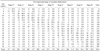
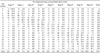
The subject population included 2490 patients between 6 and 24 years (1273 males and 1217 females, mean age: 15.7 years). Each patient's age was determined on the basis of the difference between the date of birth and the date of the X-ray. The age and gender distribution of the study population is shown in Table 1. This study evaluated 7081 third molars on 2490 panoramic radiographs (Table 2). All panoramic radiographs were taken with a Proline XC (Planmeca Co., Helsinki, Finland).
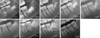
The modified Demirjian's classification system was used to evaluate the developmental status of the third molars on the panoramic radiographs, as shown in Figure 1.3,12 The assessments were carried out by one oral and maxillofacial radiologist with significant experience.
Cases of no follicles and extracted third molars were excluded from the statistical analysis. Spearman's correlation coefficients between the contralateral third molars were calculated. Descriptive statistics were obtained by calculating the means, standard deviations, and range of the chronological ages for the nine stages of third molar development. A Mann-Whitney U test was used to analyze the age differences in the developmental stages between the maxillary and the mandibular third molars and between the gender groups. A linear regression analysis was used to assess the correlation between the third molar development and chronological age. For fully developed third molars (Stage H), the probability of an individual being older than 18 years was also calculated. Statistical analyses were performed using IBM SPSS Statistics 21.0 (IBM, Armonk, NY, USA). P<0.05 was considered statistically significant.
Results
As Spearman's correlation coefficients revealed a strong correlation between the left and the right third molars, the results of both were averaged for each developmental stage.
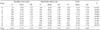
The frequency distribution of the stages for each age group is shown in Tables 3 and 4. Crypt formation of the maxillary third molars (Stage 0) was observed in two of 105 patients at age seven, and crypt formation of the mandibular third molars (Stage 0) was observed in six of 128 patients at age six. Stage A was observed from the age of 7 years and rarely beyond the age of 14 years. Crown completion (Stage D) was examined in patients aged 12 years and older. The third molars in the maxillary and mandibular arches were fully developed in 12.4% and 7.5% of the 18-year-olds, respectively, and in 98.5% and 98.7% of the 24-year-olds, respectively. Complete root formation (Stage H) in the third molars before the age of 18 years occurred in nine maxillary and mandibular third molars. The probability of an individual exhibiting Demirjian Stage H at 18 years or older was 99.1%.
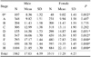
The mean developmental age and standard deviation for the modified Demirjian stages of the third molars are shown in Table 5. Initial calcification of the cusp tips (Stage A) was seen at a mean age of 9.86±1.54 years. Complete crown formation (Stage D) occurred at a mean age 14.77 ±1.68 years, and the completion of root formation without closure of the apex (Stage G) was observed at a mean age of 19.00±1.48 years. Apex closure (Stage H) occurred at a mean age of 21.96±1.70 years. The development stage of the third molars was generally more advanced in the upper than in the lower third molar. Statistically significant differences between the upper and the lower jaws were noted in Stages A-D and H. Males reached the stages earlier than females, and there were significant between-gender differences in Stages 0 and D-H(Table 6).
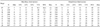
Table 7 shows the age at which each third molar development stage was observed. The third molar crypt formation was observable at as early as 7 years of age in the maxilla and 6 years in the mandible. According to our data, 75% of Stage A was visible in the upper arch in those younger than 10 years and in the lower arch in those younger than 11 years. Seventy-five percent of complete crown calcification (Stage D) was observed in the maxillary third molars in patients younger than 15 years and in the mandibular third molars in patients younger than 16 years. The minimum age at Stage H was 17 years, and 75% of those at Stage H were younger than 23 years.
A strong positive relationship was observed between the developmental stage of the third molars and chronological age. A regression analysis showed a strong correlation between age and third molar development for the maxillary (r2=0.840) and the mandibular third molars (r2=0.857).
Discussion
We investigated the development of the third molars in a 6- to 24-year-old population by using the modified Demirjian classification. In the present study, the third molars appeared at the age of 6 years and had developed completely by the age of 24 years. Developmental stages were examined at the earliest and the oldest ages. Despite wide variability, there was a clear correspondence between the developmental stage of the third molar and the age of the subject.
Crypt formation (Stage 0) was observed as early as 7 years in the maxillary third molars and 6 years in the mandibular third molars. Almost no bud formation occurred after the age of 12 years in the maxillary third molars or after the age of 13 years in the mandibular third molars, and bud formation showed wide variation between the ages of 6 and 13 years. A previous study showed that the mean age of the mandibular third molar crypt formation was 8 or 9 years, with substantial variation from about 6 to 14 years.18 Other studies reported that the crypt formation of the maxillary third molars could not be clearly seen because of overlapping adjacent structures.19,20 We found it more difficult to observe the bud formation of the maxillary third molars than that of the mandibular third molars. Therefore, the radiolucent bud of the maxillary third molar was examined at a later age than that of the mandibular third molar.
Sisman et al21 observed that the third molar began to calcify in subjects at 8 years of age. In the present study, third molar mineralization (Stage A) commenced at 7 years of age. The 9th year has been consistently reported as the mean age of the initial mineralization of the cusp tips.18 The present study found similar results.
The mean age of reaching crown completion (Stage D) was 14.77 years. Compared with reports for other populations, it was later than in an Iranian population3 and earlier than in Japanese22 or German16 populations. Bassed et al23 reported that females reached Demirjian Stage D at the latest by the age of 19 years, whereas males reached this stage at the latest by the age of 18 years. In the present study, at Stage D, the maximum age was 22 years, although crown completion was achieved in 90% of the patients by the time they were 17 years. This diversity could be explained by the differences in the selected population.
In the current study, the apical ends of the third molars were completely closed at around 22 years. This is about 1 year later than that reported in one study24 and similar to that reported in other studies.20,22,23 According to our data, Stage H was first observed at the age of 17 years, and 75% of the third molars were completed by the time the patient was 23 years.
Many studies have described the tendency of the maxillary third molar development to be more advanced than the mandibular third molar development.25,26,27 However, Olze et al28 reported no statistically significant differences in the chronology of the maxillary and the mandibular third molars. In the present study, mineralization of the maxillary third molars was more advanced than that of the mandibular third molars, and a statistically significant difference in mineralization was observed in Stages A-D and H. This finding was consistent with that reported in previous studies.25,26,27
Some studies have reported that males entered the Demirjian stages earlier than females,29,30,31 although others found no significant between-gender differences in the developmental stages of the third molars.3,7,28 In the current study, males showed earlier development than females, and there were significant gender differences in Stages D-H.
In conclusion, the present investigation could provide reference data for third molar development in the Korean population. The developmental stages of the third molars showed a strong correlation with age. The third molar developed earlier in the upper arch than in the lower arch and earlier in males than in females.


 XML Download