

 PDF
PDF ePub
ePub Citation
 Print
Print


INTRODUCTION
Food protein-induced enterocolitis syndrome (FPIES) is a non-IgE-mediated gastrointestinal food allergic disorder [1]. Diagnosis is frequently delayed because of its nonspecific symptoms and absence of classic allergic mucocutaneous or respiratory symptoms (e.g., urticaria or asthma) and a lack of definitive diagnostic biomarkers. FPIES is not easily recognized by clinicians; affected infants are often misdiagnosed and mismanaged as having acute gastroenteritis, food poisoning, sepsis, metabolic disorders, or a surgical disease [2,3,4].
In general, the diagnosis of FPIES is based on Powell's clinical criteria [5,6]; (1) exposure to the inciting food result in repetitive, profuse vomiting and lethargy, and/or diarrhea within 4 hours, without any other cause for the symptoms; (2) symptoms are limited to the gastrointestinal tract; (3) elimination of the culprit food from the diet results in resolution of symptoms; and (4) a standardized oral food challenge (OFC) test or isolated food re-introduction elicits the typical symptoms.
However, familiarity with the clinical features of FPIES in detail and awareness of the indexes of suspicion of FPIES are necessary to correctly diagnose the disease. Because of recent increasing medical concerns about FPIES and the diverse clinical observations of this hypersensitivity disorder [7,8,9,10,11,12,13], clinical terms and types of this allergic disorder have been introduced into the clinical fields. Understanding the recently defined clinical terms and subtypes of FPIES are necessary to suspect and correctly diagnose FPIES. Although physician-supervised OFC is the gold standard for a diagnosis, patients do not usually require confirmatory OFC if they have a clinical history of typical symptoms, particularly of multiple episodes [2,3,4].
The aim of this review is to provide a case-driven presentation of the terminology and clinical classification of FPIES to improve the diagnosis and management of patients with FPIES. The related literature and the author's experience are reviewed.
MAIN SUBJECTS
Clinical terms for food protein induced enterocolitis syndrome
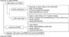
The terms for FPIES may be defined and classified according to clinical features (typical type vs. atypical type), age of symptom onset (early onset vs. late onset), duration and severity of symptoms (acute reaction vs. chronic symptom, mild to moderate reaction vs. severe reaction), and offending food (milk and/or soy FPIES vs. solid food FPIES vs. multiple food FPIES) [2,3,4].
1. According to clinical features
1) Typical (or classic) type [7] (case presentation 1-3; described in the next section as 'Clinical types of FPIES')
2. According to age of symptom onset
1) Early onset: neonate and early infancy (mostly within 3 months of age), chronically presenting FPIES when exposure to the offending food occurs continuously early in life, sepsis-like symptoms with failure to thrive, and mainly chronic symptoms [2,3,4] (case presentation 1)
2) Late onset: early and late infancy (mostly 4-7 months of age); acutely presenting FPIES after first feeding of the culprit food or after the food has been avoided from the diet and then re-introduced; repetitive and projectile vomiting with lethargic or cyanotic appearance; mainly acute reaction [2,3,4] (case presentation 2, 3)
3. According to duration and severity of symptoms
1) Acute reaction: acutely presenting FPIES; food ingested first or intermittent, milk and/or soy and/or solid foods, and mainly late onset [2,3,4] (case presentation 2, 3)
Mild to moderate symptoms during an acute reaction: few episodes of vomiting, decreased activity, self-resolving, and oral rehydration solution needed
Severe symptoms during an acute reaction: repetitive and projectile vomiting (up to 20 episodes), lethargy, cyanosis, pallor, hypotension, intravenous hydration inevitable
4. According to offending foods
1) Milk or soy FPIES: early or late onset, acute reaction or chronic symptoms, and typical type [3] (case presentation 1, 2)
2) Milk and soy FPIES: FPIES reacting to both milk and soy [3]
3) Solid food FPIES: mainly late onset; most commonly rice; and the first solids introduced into a weaning infant's diet; and rare first reaction after 1 year of age [3] (case presentation 3)
4) Multiple food FPIES: Milk and/or soy FPIES with solid food FPIES; solid food-FPIES develops in one-third of infants with milk and/or soy FPIES, and high rate of reaction to multiple foods [3]
Clinical types of food protein-induced enterocolitis syndrome
Because of increasing medical concerns and clinical observations in FPIES [7,8,9,10,11,12,13], clinical types of this allergic disorder have been defined recently and introduced into the clinical field. Understanding these terms and subtypes of FPIES is mandatory to suspect and to conclusively diagnose FPIES [2,3,4].
1. Case presentation 1
1) Case presentation
A 25-day-old male neonate was transferred to an emergency unit due to lethargy with general cyanosis since this morning. He was born at 39+3 weeks of gestation with a birth weight of 3,500 g (50th-75th percentile). He was formula-fed from birth. Beginning 7 days ago, he was presented with vomiting (2-3 times/day) and mucous and loose stool (5-6 times/day). Mild fever developed 2 days ago. Last night, weak sucking power with a sleepy condition developed. No family history of food allergy was recorded. His body weight was 2,850 g (<3rd percentile). He was drowsy and appeared malnourished and chronically ill with sepsis-like manifestation. Facial and truncal cyanosis, dried oral mucosa, and abdominal distention without hepatosplenomagaly were observed. Heart beat was weak, but without murmur, and lung sounds were relatively clear. Laboratory tests revealed peripheral blood leukocytosis with a shift to left (18% band form), thrombocytosis, hypoalbuminemia, and metobolic acidosis. Methemoglobinemia (positive ≥15%) was 25%. The results of a renal function test, serum ammonia level, serum and urine amino acid analysis, and serum immunoglobulins and T cell subsets were within normal ranges. The UniCAP test for milk was class 0. Abdominal roentgenography revealed pneumatosis intestinalis in the bowels. Gastrofiberscopy and duodenal mucosal biopsy revealed subtotal villous atrophy but without eosinophilic infiltrations. No pathogens were detected in the blood, urine, and stool cultures. The result of rectal suction biopsy was incompatible with Hirschsprung's disease. Abdominal ultrasonography showed portal venous gas. Necrotizing enterocolitis (NEC) was confirmed even though he was not a preterm baby. After methylene blue and bicarbonate injections, treatment with total parenteral nutrition, broad spectrum antibiotics, and albumin supplements was maintained for 7 days. When he was in a relatively stable condition, the open OFC test with cow's milk formula (0.15 g milk protein/kg) was performed to evaluate for possible FPIES. Baseline complete blood count with a differential was obtained before the starting of feeding. Two hours after the feeding, colic irritability with repetitive, projectile vomiting developed. Three hours of feeding, lethargy with pallor occurred, and blood pressure was 65/38 mmHg. Normal saline (30 mL/kg for 1 hour) and dextrose saline were given intravenously. Two hours after the fluid therapy, his general condition improved to a relatively active condition. Six hours after the feeding, absolute neutrophil count of peripheral blood was elevated to 5,600 cells/mm3 from baseline (positive >3,500 cells/mm3). Seven hours later, pus-like and foul-odored loose stool was observed. After 8 hours, he was fed an extensively hydrolysate formula (eHF), and no vomiting or adverse symptoms were observed. Although loose stools were observed, he recovered to be active and stable 24 hours after the OFC. He was conclusively diagnosed with FPIES [16].
2) Follow-up and resolution of FPIES
The patient was fed eHF and maintained on a strict milk protein elimination diet until 6 months of age. Then, solid foods were serially and carefully introduced under a physician's supervision, but no adverse reaction developed. At 10 months of age, OFC with soy-based formula was performed, and no reaction was observed. The eHF was changed to soy formula. At 12 months of age, OFC with cow's milk formula was performed, and repetitive vomiting and pallor occurred. At 18 months of age, he outgrew the milk intolerance and was freely fed all foods without any adverse symptoms of FPIES. Other allergic diseases (e.g., urticaria, atopic dermatitis, asthma, and allergic rhinitis) have not been observed in adolescence.
2. Case presentation 2
1) Case presentation
A 6-month-old boy, exclusively breastfed, was admitted to emergency unit with repetitive and profuse vomiting (over 10 times/hour), cyanosis, and lethargy within 4 hours after first consuming approximately 30 mL of milk formula. Four weeks previously, a similar clinical situation occurred episode after ingesting milk formula. At that time, physicians at emergency unit diagnosed him with acute viral gastroenteritis. A history revealed a common clinical feature between the two episodes; 1.5-2 hours after feeding, irritability followed by repetitive, projectile vomiting occurred. After 2 hours, a lethargic condition with ashen-gray appearance developed. Blood pressure was 65-70/42-45 mmHg. Normal saline and subsequent dextrose saline were given intravenously. Three hours after fluid therapy, his general condition improved to be relatively active. Seven hours after feeding, a mucous and bloody loose stool was observed. He was refed with breast milk, and no vomiting was observed. Twelve hours after the feeding, although a few loose stools were observed, he was completely recovered to be active and stable. The result of the UniCAP test for milk was class 0. The clinical history of two episodes with acute adverse symptoms after feeding cow's milk was compatible with the diagnostic criteria of FPIES [5,6]; therefore, he was conclusively diagnosed with FPIES.
2) Follow-up and resolution of FPIES
He was breastfed with an eHF supplement and maintained on strict milk protein avoidance until 10 months of age. Solid foods were serially introduced under a physician's supervision, but no adverse reaction occurred. At 10 months of age, OFC with soy-based formula was done and no reaction was observed. At 12 months of age, OFC with milk formula was performed and repetitive vomiting with cyanosis developed. At 15 months of age, he had outgrown the milk intolerance and could eat all foods without any adverse reactions. No other allergic diseases have been observed in adolescence.
3. Case presentation 3
1) Case presentation
A 6-month-old girl was admitted to the inpatient unit with projectile vomiting and lethargy after consuming about 20 mL of rice gruel at the start of a weaning diet. She was formula-fed from birth without any adverse symptoms. Four weeks later, an open OFC was performed with rice gruel in the inpatient unit. After feeding rice, she developed projectile vomiting, pallor with lethargy, and diarrhea. These serial symptoms and their timing were consistent with FPIES [5,6]. The result of rice-specific UniCAP test was negative.
2) Follow-up and resolution of FPIES
Thereafter, she manifested with no adverse reaction to almost all foods appropriate for her age, but after accidentally eating rice gruel at 10 months, she was presented again with symptoms of repetitive vomiting with lethargy. However, none of the allergic symptoms were demonstrated at 15 months of age after a follow-up OFC with rice gruel. No other allergic diseases have been observed in late childhood.
4. Clinical types of FPIES
Typical milk and/or soy FPIES with early onset is present with sepsis-like illness and failure to thrive (case presentation 1). Although the patient was a full-term baby, continued exposure to milk and/or soy may induce NEC or bowel perforation in severe condition. Before the time when eHF was available for clinical use in Korea (1993), four cases of NEC had developed in 10 patients who diagnosed as FPIES, even though they were all born full-term babies [21]. The differential diagnosis with infection, immune deficient disorders, metabolic disorders, or surgical diseases (e.g., Hirschsprung's disease) is extensive but necessary. This type of FPIES has rapidly decreased in incidence in Korea due to the rapid increase of breast feeding in neonate and early infancy for last 20 years (unpublished data).
Typical milk and/or soy FPIES with late onset is frequently encountered clinically and presents with repetitive vomiting, lethargy, and/or diarrhea (case presentation 2). This type is frequently misdiagnosed as viral gastroenteritis or food poisoning. After first feeding milk or soy, the observation of sequential symptoms and the timing is important to differentially diagnose FPIES. Interestingly in author's clinical experience, when changing from breast milk to infant formula (e.g., milk or soy) due to the suspicion of breast milk jaundice, a few infants firstly taking formula may present with repetitive vomiting and pallor that start within 3 hours after ingestion as acute reaction of FPIES even though they are in neonate or early infancy (i.e., acute reaction in early onset of FPIES) (unpublished data).
FPIES is elicited most commonly by milk or soy; however, rice, oats, and other solid foods may also elicit a FPIES reaction (case presentation 3). An acute reaction develops after introducing solid food into a weaning diet. This type is also frequently misdiagnosed as acute gastroenteritis. Therefore, the patients already diagnosed with milk and/or soy FPIES or even the healthy infants without FPIES should be carefully observed when firstly introducing a solid food at weaning. About one-third of infants with milk and/or soy FPIES may develop solid foods FPIES [2,3,4].
Because a FPIES is non-IgE-mediated food allergy, skin prick testing and food specific serum IgE levels to the inciting food are typically negative at the FPIES diagnosis. However, there are reports in which patients had detectable IgE of the causal food as so-called atypical FPIES [7]. Atypical FPIES has also been described in older children [14] and even in adults [15], usually related to fish and shellfish ingestion. Moreover, author and colleagues recently experienced a case of probiotic Saccharomyces boulardii-induced FPIES [18].
CONCLUSION
FPIES is an under-recognized non-IgE-mediated gastrointestinal food allergy. Diagnosis is based on clinical history, symptoms, and timing, exclusion of other causes, and if necessary, a physician-supervised OFC. Early recognition of FPIES is mandatory to prevent recurrent acute reaction with hypotension and chronic symptoms with failure to thrive. Understanding the recently defined clinical terms and classified types of FPIES is necessary to suspect and diagnose the FPIES. It is important for primary care providers and clinicians to keep FPIES in the differential diagnosis when infants present with single or multiple episodes of repetitive, projectile vomiting with lethargy or sepsis-like symptoms with failure to thrive.
 XML Download