

 PDF
PDF ePub
ePub Citation
 Print
Print


Introduction
From 2002 onward, Food and Drug Administration (FDA) warned crunchy, fried strips of potato consumers from excess use as it contained carcinogen material [1]. Frying, roasting, or baking of foods at temperatures above 120℃ resulted in a formation of a high amount of acrylamide's toxic compound [2]. Acrylamide has multiple chemicals and industrial applications [3]. These included gel electrophoresis, papermaking, and the manufacture of permanent press fabrics, manufacture of dyes and other monomers [4]. Infants and small children are more exposed to acrylamide toxicity. The toxicity is due to low body weight and high consumption of snacks (their acrylamide intake is estimated to be 2- to 3-fold higher than adults) [5]. Dietary acrylamide intake caused reproductive toxicity [6], genotoxicity, and neurotoxicity [7]. It may increase the risks of kidney and breast cancer [8]. Neurotoxic effects of acrylamide have been established in humans and animals [9]. Many researchers proved the effect of high-dose acrylamide exposure on the development of the nervous system; however, few study proved the effect of it on the development of the spinal cord [10]. There are several reports that antioxidant agents could rescue neurotoxicity induced by acrylamide via increasing antioxidant activity [11]. Rosemary (Rosmarinus officinalis) is a herb, composed of dried leaves and flowers, commonly used as a spice and flavouring agents in food processing for its desirable flavor [12]. It has anti-inflammatory [13] and antispasmodic [14] effects. In addition, it has antimicrobial [12] and antitumor [15] activities. Carnosic acid, a rosemary phenolic component has been protecting cortical neurons from glutamate and the brain from middle cerebral artery occlusion/reperfusion injury [16]. Rosemary extracts exhibited very high antioxidant activity, almost equal to that of synthetic antioxidants [17]. The main objective of this work was to address the oxidative stress of acrylamide on the development of the spinal cord motor neurons in new-born rats and protective role rosemary as antioxidant.
MATERIALS AND METHODS
Chemicals
Acrylamide was obtained from a product of leucon Sto SPP (Sigma-Aldrich, St. Louis, MO, USA). It was available in the form of white powder (99% purity) and dissolved in 1 ml distilled water [18]. It was given to rat as 10 mg/kg/day (i.e., 2 mg; 0.2 ml/rat) by oral gavage [19].
Rosemary was obtained from a local market as green leaves. The air dried leaves were powdered. Ten grams of dried plants was dissolved in 100 ml of distilled water after boiling for 5 minutes. After cooling and passing through filter paper, a clear solution was obtained [20]. During 24 hours of preparation, the extract was given to rats as (220 mg/kg, i.e., 44 mg; 0.44 ml/rat) by oral gavage twice weekly [21].
Animals
Twenty-five sexually mature female albino rats and five male albino rats (for mating) of Sprague-Dawley strain, weighing between 200–250 g (8–9 months of age) were obtained from El Helw animal house, Tanta, Egypt. The rats will be kept in cages with particular care and hygiene, artificial light/dark cycle 12 hours, at room temperature (25±2℃) and standard laboratory chow and water ad labium. The procedure approved by the ethics committee on the animal experiment of the Faculty of Medicine, Menoufia University according to the international regulations on care and use of laboratory animals. Each five of them were housed overnight with a sexually mature male albino rat for mating, and every morning vaginal smears were taken and microscopically examined for the presence of sperms. The first day of gestation was corresponding to the detection of sperms in the smears.
144 new born babies were labelled into four groups as follows.
Group I (control group): pregnant rats were given saline (normal group). The new-born were sacrificed at the age of 1-, 7-, 14-, 21-, and 42-day postnatal (6 rats each) and this corresponding to subgroups Ia, Ib, Ic, Id, and Ie, respectively.
Group II (rosemary group): pregnant rats were administered a rosemary from day 7 of gestation until day 28 after birth. The new-born were sacrificed at the age of 1-, 7-, 14-, 21-day postnatal (6 rats each) and this corresponding to subgroups IIa, IIb, IIc, and IId, respectively.
Group III (acrylamide group): pregnant rats were administered acrylamide from day 7 of gestation until day 28 after birth. The new-born were sacrificed at the age of 1-, 7-, 14-, and 21-day postnatal (6 rats each) and this corresponding to subgroups IIIa, IIIb, IIIc, and IIId, respectively.
Group IV (protected group): pregnant rats were administered acrylamide and rosemary from day 7 of gestation until day 21 after birth. The newborn were sacrificed at the age of 1-, 7-, 14-, and 21-day postnatal (6 rats each) and this corresponding to subgroups IVa, IVb, IVc, and IVd, respectively.
Group V (recovery group): pregnant rats were administered acrylamide and rosemary from day 7 of gestation until day 21 after birth. After weaning 6 rats new-borns were separated from their mothers and allowed for free water untill the age of 42 days.
Postnatal investigations
The newborns were investigated by the experimenter, and the following notes were recorded in each group (6 newborns).
Light microscopic study
At the end of each detected period. The rats were anesthetized by diethyl ether inhalation. Lumbar segment of the spinal cord was fixed in 10% buffered formalin (pH 7.4) for 48 hours. The tissue was dehydrated in ascending ethyl alcohol followed by two changes of xylene. The tissue was impregnated in paraffin wax and then embedded in paraffin wax. Sections (5 µm) were cut, dewaxed, hydrated and stained with hematoxylin and eosin, silver and toluidine blue stain [24].
Immunohistochemical study
For the immunohistochemical study, the spinal cord paraffin sections were deparaffinized and rehydrated in descending grades of alcohol. Following blocking of endogenous peroxidase activity with 3% H2O2 in methanol and nonspecific binding sites with a protein blocker, the primary antibody (1:500, neurofilament [NF]; 1:300, myelin basic protein; Abcam, Cambridge, MA, USA) added with overnight incubation in a cold room. On day 2, the biotinylated secondary antibody (Vector, Peterborough, UK) was added at a concentration of 2% for 30 minutes (37℃) followed by addition of the avidin-biotin-peroxidase complex (Vector). All steps were performed at room temperature in a humidity chamber [24].
Biochemical assay
For determination of antioxidant enzymes, segments from lumbar part of the spinal cord were removed and homogenized in potassium phosphate buffer solutions (50 mM, pH 7.5) using a Potter-Elvehiem homogenizer to give a 10% homogenate. Homogenates were centrifuged at 1,500 ×g for 10 minutes at 4℃; the supernatant was recovered, placed on ice, and immediately used for the determination of peroxidase and superoxide dismutase (SOD). The activity of SOD was determined calorimetrically according to the method of Marklund and Marklund [25]. While peroxidase activity was determined according to the method of Kar and Mishra [26].
Molecular study: Comet assay for motor neuron cell [27]
Preparation of motor neuron cell suspensions:
Spinal cords were isolated from rats that were anesthetized deeply with a mixture of enflurane:oxygen:nitrous oxide (1:33:66) and then decapitated. After removal of the pia matter, lumbar/cervical enlargements were dissected segmentally under a surgical microscope and then the segments were microdissected into gray matter columns of the ventral horn without appreciable contamination of dorsal horn and surrounding white matter funiculi. Gray matter tissue columns from spinal cord were collected and rinsed in a cell culture dish on ice containing dissection medium (1Ca2+ and Mg2+-free Hanks balanced salt solution [Gibco BRL, Grand Island, NY, USA] supplemented with glucose and sucrose). These tissues were used to prepare motor neuron cell suspensions.
Encapsulation:
A volume of 50 µl cell suspension (containing ~4.4×10⁴ motor neurons) was added to 200 ml 0.7% low melting point agarose at 4℃, and then layered onto a pre-coated microscopic slide with 100 µl of low-melting-point agarose and covered with a coverslip. The agarose was gelled at 4℃, and then the coverslip was removed. The slides were immersed in lysing solution (2.5 M NaCl, 100 mM EDTA, 10 mM Tris-HCl buffer, pH 10, 1% sodium sarcosinate with 1% Triton X-100 and 10% DMSO; Sigma-Aldrich) for ~1 hour.
Electrophoresis:
The slides were washed with distilled water to remove all salts and then placed in a horizontal gel electrophoresis unit (CBS Scientific, San Diego, CA, USA) filled with fresh electrophoretic buffer (1 mM disodium EDTA and 200 mM NaOH, pH 13).
Electrophoresis was conducted for 20 minutes at 25 V and 300 mA. Slides were then stained with ethidium bromide (Sigma-Aldrich). Slides were examined with a Carl Zeiss fluorescent microscope (Jena, Germany) equipped with a 510 nm excitation filter and a barrier filter of 590 nm. In damaged cells, breaks appear as fluorescent tails extending from the core towards the anode. The tail length reflects the amount of DNA breakage in the cell [28].
Quantitative study
By using Image analyzer software (Image J 1.47v, National Institute of Health, Bethesda, MD, USA) (Department of Anatomy and Embryology, Faculty of Medicine, Menoufia University) the following parameters were calculated.
(1) Number of motor neurons and the number of neuroglia cells (the neurons were differentiated from glial cells by glial nuclei diameter <5 µm).
(2) Color intensity of Nissl granules and the surface area of the brown color of NF and myelin basic protein (MBP) immunohistochemistry.
(3) The migrated nuclear DNA was considered as a damaged DNA spot. The migration was evaluated by measuring the basal nuclear DNA and migrating DNA. In 50 randomly selected cells per sample, the used comet parameters for the evaluation are tail length, tail DNA% and tail moment. The tail length was measured from the center of the nucleus towards the end of the tail, the percentage of DNA in the tail DNA% and tail moment (Tail moment=Tail length×Tail DNA%) [29].
Statistical analysis
SPSS version 20 (IBM Co., Armonk, NY, USA) was used for the statistical analyzes. The data were analyzed using Mann-Whitney test and Kruskal-Wallis test followed by Post hoc test to compare various groups with each other. Results were expressed as mean±SD. The level of significance was expressed as P>0.05 for insignificantly [30].
RESULTS
General developmental observations
Signs of acrylamide toxicity were observed postnatally in the treated mothers. It represented by ataxia, splayed hind limbs, weakness of the hind limb muscles, and paralysis. The limb weakness decreased both food and water consumption. There were no significant changes in all tested parameters of the offspring of control/rosemary groups. Neither congenital anomalies nor deaths were reported in-between offspring.
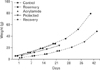
Body weight:
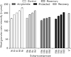
There was a significant increase in body weight with a progression of age (P<0.05). In an acrylamide-treated group, the increase in body weight was significantly lower than a control group (P<0.05). While supplementation of aqueous rosemary extract with acrylamide led to an extremely significant increase in body weight with age (P<0.001) when compared with acrylamide-treated rats. Also, acrylamide withdrawal resulted in significant increase in body weight (P<0.05). However, it was still significantly lower than the control group (Fig. 1).
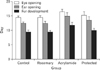
Developmental landmarks:
At birth, the newborns of all groups were hairless. The time of fur appearing and both ear and eye opening were significance retarded in the acrylamide-treated group (P<0.05) when compared with that of the control group. In the protected group, the developmental parameters showed significantly earlier development (P<0.05) when compared with an acrylamide-treated group (Fig. 2).
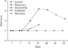
Gait scores:
At birth, all rats were unable to walk except at the age of the 12–13 day postnatal. Rats of the acrylamide-treated group showed a significant increase in gait score from day 14 to day 21 (P<0.05) when compared with control group. However, this change significantly decreased (P<0.05) when rats treated by rosemary with acrylamide, it was significantly low at day 14. Also, withdrawal of acrylamide for three weeks led to a decline in gait score but still significantly higher when compared with control group (Fig. 3).
Skeletal landmarks:
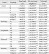
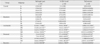
Both control and rosemary groups showed a steady increase in head length, cervical rump length and tail length with age. In acrylamide group, the head length of acrylamide rats significantly decrease (P<0.05) than the control group at the age of 14 days. The cervical rump was significantly decrease (P<0.05) from control group from day one to day 14. It became pronounced at day 21 (P<0.001) and tail length significantly decrease (P<0.05) at day 21. Rosemary supplementation was significantly increase (P<0.05) in the tail length cervical rump in acrylamide rats at day 1 and that increase (P<0.001) from day 7 inward. Also, withdrawal of acrylamide for three weeks led to increasing in three parameters but still significantly lower when compared with control group (Table 2).
Tissue biochemical results
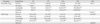
Both control and rosemary treated animals showed no significant differences (P>0.05). With the advancement of age, both groups showed a significant decrease in the spinal levels of an antioxidant marker (SOD) and increased in the content of peroxidase as compared with controls (P<0.05).
With the advancement of age and compared with controls group, the acrylamide-treated rats showed a significant decrease in the spinal levels of an antioxidant marker (SOD) and an important increase in the content of peroxidase (P<0.001). Rosemary administration to acrylamide-treated (protected group) rats resulted in a significant rise in SOD and a significant decrease in the peroxidase level when compared with acrylamide-treated rats (P<0.001). Withdrawal of acrylamide (recovery group) ameliorated both parameters but still a significant difference from control (P<0.001) (Table 3).
Histopathological results
Control and rosemary treated groups:
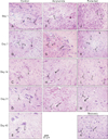
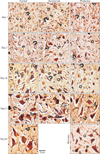
At birth, histological sections of the control, as well as the rosemary groups, revealed that anterior horn cell of the spinal cord showed numerous well-differentiated motor neurons in-between scattered small capillaries and different forms of neuroglia. With age advanced, the neurons became more basophilic. It showed a significantly increase in number and size (P<0.05) while neuroglia cells showed significantly decrease in number (P<0.05) (Table 4, Fig. 4A–E).
The motor neurons of anterior horn cells had a central vesicular nucleus with an eccentric nucleolus, partially myelinated axons with no-dendrites. The oligodendroglia, astrocyte, and microglia represented the presented neuroglia. With advanced age, the neurons acquired small dense granules, long branched dendrites, and long myelinated and nodded axons. Many microglia were reported in early age while oligodendroglia became more numerous with the advancement of age (Fig. 5A–E). With advanced age, the neurons acquired small dense granules, long branched dendrites and long myelinated and nodded axons. Many microglia were reported in early age while oligodendroglia became more numerous with the advancement of age (Fig. 5A–E).
Acrylamide group:
In comparing with control, the anterior horn cell sections at day 1 revealed non-significant decrease (P>0.05) in differentiated motor neurons number and size and significant increase (P<0.05) in neuroglia cells. The neuropil showed small vacuolation, hemorrhage, dilated congested capillaries. With the increase in age, the neurons showed a steady decrease in number and size marked at day 21 (P<0.001 and P<0.05, respectively). The neuropil showed a significant increase in neuroglia that became marked (neurogliosis) at day 21 (P<0.001). A concomitant increase in vacuolation, congestion and neuronophagia were noticed (Table 4, Fig. 4F–I).
Acrylamide rats motor neurons at day 1 showed either neurofibrillary tangle or early central chromatolysis (eccentric nucleus and homogenous cytoplasm) and segmental demyelinated, slight swollen axons. With an increase in age, the motor neuron showed extensive pathological future as: end stage chromatolysis, degenerated pyknotic neurons, attenuated dendrites, and giant swollen destructed axons with an irregular myelin pattern. A remarkable number of astrocytes, many microglia cells also noticed (Fig. 5F–I).
Protected group:
In compare with acrylamide group, rats of protected group showed, with age increase, significant increase and a decrease (P<0.05) in the number of motor neurons and neuroglia respectively starting from day 1. From day 14, the neurons size began to show significant increase (P>0.05). The architecture of ventral horn showed noticed improvement with few areas of vacuolation, vascular congestion, and few degenerated cells (Table 4, Fig. 4J–M). Some neurons showed with short dendrites, and slight swollen destructed and demyelinated axons (Fig. 5J–M).
Recovery group:
In compare with acrylamide group, the recovery rats showed significantly increased number and size of motor neurons (P<0.05) and significant decrease in neuroglia number (P<0.05). Although the general architecture showed improvement great areas of vacuolation, vascular congestion, and few degenerated cells were still observed (Table 4, Fig. 4N) and neurons with short dendrites and visible swollen destructed and demyelinated axons (Fig. 5N).
Histochemical and immunohistochemical results
Toluidine blue staining:
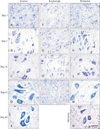
At birth, anterior horn control section stained with toluidine blue showed motor neurons with round body, defined nucleus and clear cytoplasm containing Nissl granule. The latter appears as fine scanty faint basophilic granules inside the cytoplasm and proximal part of dendrites. Both control and rosemary rats showed no significant difference (P>0.05) in Nissl granules content and intensity which increased significantly (P<0.05) with age advancement. Compared with the control group at the same age, chronic acrylamide administration showed significantly reduction of Nissl granules content and intensity at the age of 21 days (P<0.001). On day 7 and age forward, protected group showed steady significant increase in the Nissl granule content and intensity (P<0.05). Withdrawal of acrylamide for 21 days after acrylamide stop-page showed an increase in Nissl granules content and intensity but still significantly lower than the control group (P<0.05) (Figs. 6, 7).
NF immunostaining:
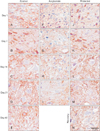
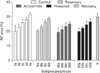
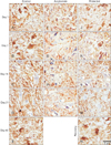
At birth, the pattern of NF expression immunohistochemistry stain expression (specific for axonal neurofilament), in the anterior horn control sections, appeared as fine scanty parallel lines. Both control and rosemary rats showed no significant difference (P>0.05) in NF protein expression (area %) which increased significantly (P<0.05) age advancement. Compared with control group, chronic acrylamide administration showed steady significant decreased in NF expression with age and became marked (P<0.001) at the age of 21 days. Compared with acrylamide group, at the same age, concomitant administration of rosemary with acrylamide significantly increase (P<0.001) in NF protein at the age of 7 days. While the withdrawal of acrylamide increased NF protein but still significantly lower (P<0.05) than a control group (Figs. 8, 9).
MBP immunostaining:
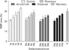
At birth, the pattern of MBP immunohistochemistry stain expression (specific for myelin sheath warping axon and oligodendroglia) in anterior horn control section appeared scanty small fine parallel brown lines or rings. Both control and rosemary rats showed no significant difference (P>0.05) in MBP content (area %) which increased significantly (P<0.05) with age advancement. Compared with the control group at the same age, chronic acrylamide administration showed significant reduction (P<0.001) of MBP content (area %) at the age of 14 days.
Compared with acrylamide group, at the same age, concomitant administration of rosemary with acrylamide significantly increase MBP content in the ventral horn (P<0.05). While acrylamide withdrawal showed an increase in MBP content but still significantly lower than the control group (P<0.05) (Figs. 10, 11).
Single comet assays result
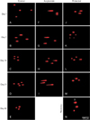
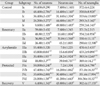
At birth, the pattern single motor neuron comet assay was detected morphologically by a faint small tail, morphometrically by few % DNA in the tail and mathematically by small tail moment value. Both control and rosemary rats showed no significant difference (P>0.05) in previous mentioned three parameters which increased significantly (P<0.05) with age advancement. Compared with control groups, acrylamide administration showed a steady, significant increase in DNA damage (P<0.001) with age. The significant increase in tail length, % DNA in tail and tail moment represented DNA damage. Compared with acrylamide group cumulative concomitant administration of rosemary significantly improved in DNA by decreasing in tail length, % DNA in tail and tail moment. This improvement was more pronounced in younger ages (P<0.001) than in older ages (P<0.05). Acrylamide withdrawal led to a reduction of DNA damage but the tail moment still significantly lower than control of the same age (P<0.05) (Table 5, Fig. 12).
DISCUSSION
Great numbers of the population are exposed to acrylamide toxicity as it formed during baking, grilling, or frying of starchy foods [8]. Although the oxidative stress of acrylamide in the central nervous systems has been reported [31], its effect on the neurons developments takes little attention in spite of its crossing the placental barrier [3233] and expressed in milk during lactation [34]. As a powerful phenolic natural agent, rosemary possessing a protective activity on nervous system [16]. In this study, we investigated the effect of acrylamide on postnatal development of spinal cord motor neurons in new born rats and the possible protective effect of rosemary.
In this study, the maternal acrylamide exposure during the gestation and lactation periods proved its toxic developmental effects, as it delayed fur appearing and ear and eye opening [35]. In addition to its potential teratogenic effects, as it and decreased head, crown-rump, and tail lengths [36].
As reported in previous studies [3738], our study reported important clinical signs in acrylamide intoxicated rat. This was represented by a decrease in body weight and gait disorder. The decrease in the body weight might be due to losing of appetite resulted from leptin transport disorders, accompanied acrylamide toxicity [39] as well as reduction of prolactin of affected mother [40]. While gait disorder might be due to the development of neuropathic syndrome (ataxia and dragging of hind limb) that recorded to acrylamide poison. In our work, this could be supported through (1) histopathological changes, neural axon and myelin sheath degenerations and accumulation of the neurofilament, (2) biochemical increase in oxidation enzyme [41].
The obvious toxic effect of acrylamide on motor neuron proved in our study (undifferentiation, degeneration, and chromatolysis) might also explain the increased gait score and the retardation of sensorimotor reflexes as observed by others [4243]. As motor activity regulated by the functional integration of neuronal activity in various regions of the brain and the spinal cord [44].
In acrylamide group, the chronic destructive effect on new born neuronal axons that accompanied with decreased myelin and NF protein were in agreeing with Fleck [45]. Also, it disagrees with Takahashi et al. [46] who didn't observe acrylamide developmental neural toxicity in sciatic nerve of a new born of a perinatal exposed mother. They assumed this to the high plasticity of the nervous tissue and decrease toxic through decrease milk of the mother. This discrepancy might be due to the different animal species and different plasticity of central from the peripheral nervous system.
In this study, we also approved the apoptotic effect of acrylamide on developing motor neurons in the form of central chromatolysis and significant DNA destruction by comet assay. This might be due to the high-affinity acrylamide metabolic glycinamide derivative to form DNA adducts [47].
The destructive effect acrylamide on RNA and DNA added a more explanation to increased score gait by a decrease in new-born's activity through impairing neural protein production as mentioned by others [4048]. This manifested in our work through a significant decrease in intracellular Nissl granules.
In acrylamide group, a concomitant significant decline of antioxidant enzymes with the developing spinal cord tissue damage proved oxidative stress of acrylamide substance. This recorded by others [43] who attributed its oxidation effect to change in the neuron cytoskeleton or its necrotic membrane effect and mitochondrial dysfunction. This developmental acrylamide motor neuron toxicity could be cleared by known its mode of action as previously mentioned [49]. They said: as a soft electrophile acrylamide acts by forming Michael adducts with soft nucleophilic sulfhydryl thiolate sites on proteins. In our work, this could be obvious in nerve terminal protein targets in this work: NF and myelin protein. The change in the latter might be relevant to the mechanisms of the neuro-filamentous axonopathies induced by acrylamide as proved by others [50]. Wei et al. [51] added the changes of calpain activity as a cause of axonopathy produced by acrylamide. Collectively, rapidly absorption and distribution of acrylamide through the tissue [52] might explain its multi-toxicity observed in this study.
Our result revealed that rosemary extract was able to act as a neuroprotective agent against acrylamide-induced motor neuron toxicity not only on the through biochemical antioxidant effect as well as histomorphological protection effect and anti-apoptotic effect. A significant enhancement of performance rosemary on central nervous system has been proved by others [53]. Rosemary exerted protective effects against acrylamide-induced oxidative damage via its detected antioxidant properties and decreased lipid peroxidation and hence tissue damage. Rosemary might exert its antioxidant through the ability to protect cell membranes against attack by reactive species through its high concentration of carnosic and rosmarinic acid recognized as natural antioxidants. Carnosic acid as phytopolyphenol can trap oxygen/nitrogen-based free radicals. However, as a polyphenol, carnosic acid has several enol sites which can ionize to a nucleophilic enolate that can scavenge electrophiles such as acrylamide and the unsaturated aldehydes (e.g., acrolein, 4-hydroxy-2-nonenal) that mediate oxidative stress [54].
A significant proliferation of neurons detected in the protected group might occur due to rosemary neural proliferative effect [55]. The regeneration of axon accompanied with an increase in NF and myelin protein might be due to the enhancement effect of the rosemary extract constitution, carnosic acid and carnosol, on nerve growth [15]. As rosemary, phenolic diterpene produces enhancement of reperfusion circulation injury [16] and prevents acetylcholine breakdown that enhances transmission [56]. The recorded improvement in the recovery group comes in line with others [57] who observed human neural recovery from acrylamide after several months to a year of cessation of exposure. The absence of postnatal maternal acrylamide exposure might play a role. As well as, the catalyzed of its glutathione conjugation (N-acetyl-S-cysteine) in the liver, brain, and skin both enzymatically and non-enzymatically and its short half-life span, as it eliminated in rats urine after about two hours in rats [58]. Smaller amounts eliminated via faeces and exhaled CO2 [59].
From the results of the present work, we can infer that acrylamide has deleterious effects on the postnatal development of spinal cord motor neurons as clarified by biochemical, histopathological, immunohistochemical, and molecular changes. Also, rosemary was found to have a protective effect against acrylamide toxicity. Therefore, we highly recommended rosemary as a promising neuroprotective natural agent especially when added to starch frying food.
 XML Download