

 PDF
PDF ePub
ePub Citation
 Print
Print


Introduction
The cecum and appendix are the initial parts of the large intestine and are situated in the right iliac fossa. The appendix is a narrow wormlike structure arising from the posteromedial wall of the cecum about 2 cm below the ileocecal junction and is not constant in its position. The length of the appendix varies from 2 to 20 cm (average, 2 to 9 cm) [1]. Acute appendicitis is one of the most common diseases and is easy to diagnose [2]. However, when the appendix is situated in an abnormal position, the diagnosis of acute appendicitis becomes difficult [3]. Delayed diagnosis or misdiagnosis of subhepatic appendicitis might lead to perforations of the appendix, which is a clinical emergency [4]. One of the early cases of subhepatic appendicitis was reported by King in 1955 [5]. The terminal part of the ileum joins to the left side of cecum in the right inguinal region [6]. When the cecum is in the subhepatic position, the terminal part of the ileum passes through the right iliac fossa and joins the cecum at different angles [7]. In this report, we discuss the possible complications resulting from a subhepatic appendix and a retroperitoneal ileum.
Case Report
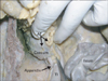
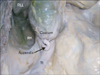
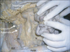
During dissection classes for undergraduate medical students, the subhepatic position of the cecum and vermiform appendix was noted in an adult male cadaver aged approximately 70 years. The cause of death was natural, and there were no other visible abnormalities in the body. The cecum was of the "ampullary type," and the vermiform appendix was attached to its posteromedial side. The appendix was uncinate and made a "U"-shaped bend, and its tip was located in the 11 o'clock position (Figs. 1, 2). The cecum had appendices epiploicae, and the terminal part of the ileum was retroperitoneal (Figs. 2, 3). The cecum and appendix were closely related to the gall bladder, the right lobe of the liver, and the right kidney. The ascending colon was very short. The mesentery of the small intestine ended at the right iliac region, and the terminal, retroperitoneal part of ileum ascended vertically to the cecum from the right iliac fossa (Fig. 3). The retroperitoneal ileum was approximately 30 cm in length and opened into the cecum at approximately a 170° vertical axis to the cecum.
Discussion
During the development of the intestine, the vermiform appendix and cecum develop from the cecal bud of the postarterial segment of the midgut loop. In the initial stages of development, the cecum and the appendix are of the same diameter, but later, the appendix narrows down because of the faster growth of the proximal part of the cecal bud. The midgut herniates to the umbilical cord as a part of the physiological umbilical hernia in the sixth week of intrauterine life, and returns back to the abdomen in the tenth week of intrauterine life. When the intestine returns to the abdomen, the cecum and appendix occupy the subhepatic position. Later, in the eleventh week, they descend to the right iliac fossa [8, 9]. At birth, the appendix is attached to the tip of the conical cecum, and this condition rarely persists into adulthood. In this case, the terminal part of the ileum would have lost its mesentery through zygosis around the tenth week of intrauterine life, as soon as the intestine returned to the abdominal cavity. This, in turn, would have prevented the cecum from descending to the ileac fossa. However, in the current case, the cecum had grown into the ampullary type from its infantile type in the subhepatic region. The cecum and appendix usually do not possess appendices epiploicae, but in our case, the cecum had appendices epiploicae. During our literature survey, we could not come across any such case of the cecum with appendices epiploicae. Subhepatic acute appendicitis might lead to many problems. It might infect the gall bladder and bring about symptoms of cholecystitis and cholelithiasis, or it might even mimic liver abscess [4, 10].
In the current case, the cecum and appendix were closely related to the right lobe of the liver, gall bladder, and right kidney. The appendix was sandwiched between the cecum and the kidney. Acute appendicitis in this case might spread to the right kidney, suprarenal region, and liver, and can lead to a diagnostic dilemma. The knowledge of this type of variation is of utmost importance to radiologists and surgeons. The retroperitoneal part of the ileum might have been mistaken for the ascending colon because it occupied the area that was supposed to be occupied by the ascending colon. This might lead to confusion in the interpretation of barium enema and computed tomographic scan findings. The pus from the perforated appendix might enter the lesser sac easily in this case. Additionally, the fixation of the terminal part of the ileum might limit its peristaltic movements. The knowledge of these concurrent variations might be very useful to clinicians of various medical disciplines.
 XML Download