

 PDF
PDF ePub
ePub Citation
 Print
Print


Congenital left atrial band (CLAB) is a fibromuscular band observed in the left atrium and observed in 2% of necropsy.1) However, the clinical significance has not yet clearly identified and clinically diagnosed cases are very rare.2) Histopatholgic study showed that the anomalous bands were composed of fibrous and muscular tissues without Purkinje cells.3) The fibromuscular bands of the left ventricle or right atrium have been reported to be associated with specific types of tachycardia. CLAB has also been reported to raise the incidence of supraventricular arrhythmia.1)
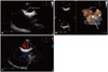
A 76-year-old man with persistent atrial fibrillation was admitted to the hospital for elective electric cardioversion. He had a history of type 2 diabetes and dyslipidemia without other underlying diseases. His transthoracic echocardiogram was unremarkable and transesophageal echocardiography (TEE) was performed for screening intracardiac thrombus before electric cardioversion. In TEE, non-mobile linear structures attached from the interatrial septum in the left atrium were observed (Fig. 1A and B) and there was no flow limitation or acceleration in the color Doppler study (Fig. 1C, Movie 1). There were no significant valvular abnormalities of all cardiac valves. Because flow acceleration in the Doppler study was not observed and three-dimensional imaging study shows a band-like linear structure rather than a membrane, we considered this linear structure as CLAB. Electric cardioversion successfully converted atrial fibrillation to normal sinus rhythm without complication.
There is a case report of two patients with CLAB and cryptogenic stroke suggesting that CLAB could be considered a potential cause of a cardioembolic event.4) Therefore, although the association between atrial fibrillation and CLAB is not clearly identified, there is a possibility that atrial fibrillation could be induced from CLAB. However, according to a case report by Uutake et al.5) in 2015, electrophysiological study failed potential arrhythmogenic activity of CLAB. Therefore, additional studies on the association of CLAB with atrial fibrillation are needed. In this report, we present the characteristic features of two-dimensional and three-dimensional echocardiograms of CLAB.
 XML Download