

 PDF
PDF ePub
ePub Citation
 Print
Print


Introduction
The detection of right ventricular (RV) systolic dysfunction (RVSD) due to pulmonary vascular disease in preterm children can be critical for determining prognosis.1) Late-onset pulmonary hypertension (PH) has been reported in extremely low birth weight infants2) and children with no or mild bronchopulmonary dysplasia (BPD),3) advocating the need for long-term echocardiographic screening in preterm children. A subset of preterm infants without BPD but with PH detected at 36 weeks of postmenstrual age showed altered RV function on deformation imaging, which persisted up to 1-year corrected age.4) Thus, primary vascular injuries of the lung may cause RV dysfunction even in preterm infants, independent of BPD.5) To date, the sequential assessment of RV function beyond 1-year corrected age has not been performed in preterm children; therefore, the progress of potential RV dysfunction beyond this period in preterm children remains unclear.
The detection of RVSD due to pulmonary vascular disease in preterm children can be challenging because even in patients with significantly elevated pulmonary artery pressures, symptoms may be subtle.6) In the RV, the longitudinal shortening of myocardial fibers contributes mainly to stroke volume during systole.7) Therefore, measuring decreases in RV strain in the longitudinal direction in the presence of afterloads, such as increased pulmonary vascular resistance, would enable the sensitive detection of RVSD in preterm children.8) Myocardial strain, indicating changes in the length of myocardial fibers,9) is more sensitive in detecting early myocardial changes among preterm infants than are conventional echocardiographic parameters.10) We speculated that strain parameters could sensitively detect the progression of potential RVSD among preterm children. Velocity vector imaging (VVI) utilizes speckle and endocardial contour tracking to assess regional and global cardiac function.11) Accordingly, we used VVI to detect possible RVSD in asymptomatic preterm children from infancy to 24-month corrected age, based on myocardial deformation parameters [namely, RV longitudinal peak systolic strain (LPSS)] and to investigate the possible clinical parameters correlated with RV LPSS.
Methods
This study was approved by the Institutional Review Board of our institution (approval number 2015-09-159-002).
Study population
Preterm infants with < 33 weeks of gestational age (GA) who were admitted to the neonatal intensive care unit of CHA Bundang Medical Center were identified from our medical database and retrospectively studied. Infants with structural congenital heart diseases, except for patent ductus arteriosus (PDA) and patent foramen ovale, and those with genetic disorders were excluded. Demographic information, including GA, birth weight, age, weight, and heart rate during echocardiography, and systolic and diastolic blood pressures were collected. Patient age was specified as corrected age, which was obtained by subtracting the number of weeks born before 40 weeks of gestation from the chronological age.12)
Characteristics during the antenatal and perinatal periods, duration of mechanical ventilation, history of BPD, and history of hemodynamically significant PDA (hsPDA) were assessed. BPD was defined as the requirement for supplemental oxygen at 36 weeks of GA.13) hsPDA was PDA that required medical intervention. Term infants referred to our clinic for murmurs who were without significant structural heart diseases other than small patent foramen ovale were studied as controls.
Echocardiographic assessment
We retrospectively reviewed the echocardiograms performed at a mean of 4-month corrected age (first exam) and 24-month corrected age (second exam) using commercially available ultrasound equipment (Acuson SC 2000, Siemens Medical, Mountain View, CA, USA).
All echocardiograms were performed as recommended by the American Society of Echocardiography.14) Conventional echocardiographic parameters were measured as recommended.15) PDA diameter was measured at the pulmonary end. Because we excluded patients with significant pulmonary stenosis, maximal tricuspid regurgitation (TR) jet velocity from continuous- wave Doppler and right ventricular systolic pressure (RVSP) derived from the modified Bernoulli equation (TR jet velocity2 × 4) was obtained.6)16) We determined PH based on the presence of findings (primary echocardiographic criteria for PH) of RVSP > 40 mm Hg, RVSP/systemic systolic blood pressure > 0.5, and cardiac shunt with bidirectional or right-to-left shunt or any degree of interventricular septal flattening.3)
Left ventricular ejection fraction, fractional area change (FAC) of RV, expressed as a percentage change in the RV chamber area at end-diastole and end-systole,1) tricuspid inflow Doppler velocity during early diastole (E), tricuspid annular tissue Doppler velocity during early diastole (E′), and the ratio of tricuspid E/E′ was obtained according to previous recommendations.17) Isovolumic contraction time (IVCT) and isovolumic relaxation time (IVRT) were obtained by Doppler tissue imaging, and myocardial performance index was calculated as follows18): RV myocardial performance index = (IVCT + IVRT)/RV systolic ejection time.
VVI analysis
Digital echocardiographic images of the study population were obtained at 70 frames per second and stored for subsequent offline analysis. On average, three cardiac cycles were studied. All offline analyses were performed using vendor-customized VVI software (Siemens Medical, version 3.0). Two independent investigators who were blinded to the clinical data of the study population at the time of analysis performed the VVI analysis.
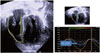
In the apical four-chamber view, at the onset of the QRS wave, we manually tracked the endocardial border of the RV using the VVI software to acquire RV LPSS as recommended.20) The RV LPSS was identified as the highest point of the average strain and strain rate curve derived from six segments (three from the RV free wall and three from the interventricular septum) (Fig. 1).20)
Statistical analysis
All values are expressed as mean ± standard deviation.
SPSS version 24 (IBM SPSS Statistics 24, IBM Corp., Armonk, NY, USA) and MedCalc for Windows version 17.5 (MedCalc Software, Ostend, Belgium) were used to analyze data. Echocardiographic and clinical data between the two groups were compared using Student's t-test or Mann-Whitney U-test, as appropriate. A p value of < 0.05 was considered significant. Pearson's or Spearman's correlation was performed, as appropriate, to determine correlations. One observer performed an offline repeat analysis of RV LPSS in 15 subjects at 4 weeks apart to determine intraobserver variability. Two independent observers who were blinded to clinical data of the children at the time of analysis performed a separate offline analysis of RV LPSS in 15 children to determine interobserver variability. The mean percentage error, which were obtained by calculating the absolute difference of the two datasets divided by the mean of the two datasets,21) were used to determine intraobserver and interobserver variabilities.
Results
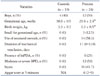
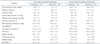
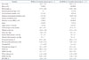
A total of 34 children (preterm children, n = 24; controls, n = 10) were studied. Demographic and clinical data of preterm children and controls are shown in Table 1. Median GA of the preterm children was 29.55 weeks (range: 24–33 weeks GA). All preterm children were asymptomatic at both time periods, and no patients needed oxygen or demonstrated hsPDA at both time periods. Echocardiographic data of preterm children and controls are shown in Table 2. Among conventional echocardiographic parameters, compared with controls, preterm children showed increased mean tricuspid E/E′ during the first exam (preterm children vs. controls, 4.2 ± 1.6 vs. 3.2 ± 1.1; p < 0.05) and showed increased mean TR jet velocity during the second exam (preterm children vs. controls, 1.3 ± 0.6 vs. 0.8 ± 0.5 m/s; p < 0.05). Two preterm children showed interventricular septum flattening at the first exam and were determined to have PH, but neither were shown to have PH at the second exam. Over time, in preterm children, the mean heart rate and mean TR jet velocity decreased significantly, and the mean PAAT increased significantly. The mean PAAT did not differ between preterm children and controls during the exam. The mean RVSP and MPAP of the preterm children were < 40 mm Hg at both time periods. The mean MPAP decreased over time in preterm children (first exam vs. second exam, 31.1 ± 4.2 vs. 24.3 ± 8.0 mm Hg; p < 0.05), although the mean RVSP remained elevated compared with the controls (preterm children vs. controls, 8.0 ± 7.0 vs. 3.9 ± 4.9 mm Hg; p < 0.05) during the second exam.
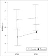
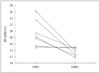
Over time, in preterm children, the mean RV LPSS did not show significant difference between both exams; however, the mean RV LPSS was decreased in preterm children compared with that in controls at both time periods (Fig. 2). We further analyzed the change of RV LPSS in individual preterm children at both time periods. The change over time of RV LPSS in individual preterm children who showed RVSD at 24 months corrected age is shown in Fig. 3. We defined decreased RV LPSS of < 16% as RVSD (referencing the lower level of RV LPSS of the controls in our study). In 11 preterm children (45.8%), RVSD was detected during the exam. In 8 of these 11 children, RVSD improved during the second exam. In 3 of the 11 preterm children with RVSD during the first exam, RVSD was persistently detected during the second exam. In five preterm children (20.8%), RVSD was not detected during the first exam, but progression to RVSD was noted during the second exam. Totally, in seven preterm children (29.2%), RVSD was detected at 24-month corrected age. The demographic, clinical, and echocardiographic data of preterm children with and without RVSD at 24-month CA are shown in Table 3.
Intraobserver and interobserver variability of RV deformation indices
The mean percentage error for intraobserver and interobserver variabilities in RV LPSS was 20% and 21%, respectively.
Correlation of RV LPSS with clinical variables
In preterm children, small birth weight was negatively correlated significantly with RV LPSS during the first exam (r = -0.446, p = 0.029). GA, sex, systolic and diastolic blood pressures, history of severity of BPD, presence of hsPDA, use of antenatal steroids, and duration of mechanical ventilation all did not show a significant correlation with RV LPSS in preterm children at both time periods.
Discussion
In our study, the overall mean RV LPSS of preterm children showed no significant change from a mean 4–24-month corrected age over time but remained decreased at both time periods compared with controls. The mean TR jet velocity decreased over time in preterm children but remained increased compared with controls at mean 24-month corrected age. In seven of 24 preterm children (29.2%), RVSD, defined as RV LPSS < 16%, was detected at a mean of 24-month corrected age. In five of seven children with RVSD at 24-month corrected age (20.8% of all preterm children), no RVSD was detected at mean 4-month corrected age, and RVSD progressed after a mean of 4-month corrected age. Low birth weight was negatively correlated with RV LPSS in preterm children at a mean of 4-month corrected age; however, the severity of BPD, duration of mechanical ventilation, presence of hsPDA, and use of antenatal steroids all did not correlate with RV LPSS. Age and weight during the exam did not correlate with RV LPSS in preterm children at both time periods.
Our study is the first to sequentially analyze RV LPSS in preterm children from a mean 4–24-month corrected age. Our results of the mean RV LPSS remaining decreased compared with controls from 4- to 24-month corrected age may be due to elevated pulmonary vascular resistance in the preterm children compared with controls, as TR jet velocity was elevated in the preterm children compared with controls at 24-month corrected age.
However, TR jet velocity, in contrast to RV LPSS, was not significantly different between preterm children and controls at 4 months corrected age in our study. Since detection of TR jet velocity has not always been possible for children with chronic lung disease,6) RV LPSS might be a more sensitive parameter of RV dysfunction due to potentially elevated pulmonary vascular resistance in preterm children. In our study, during the second exam, RVSD was detected in 7 of 24 preterm children (29.2%). In these children, RVSP was significantly higher compared with the controls during the second exam, although not reaching PH levels. As only two of these seven children had a history of moderate or severe BPD, we speculate that in at least five of these children primary lung vascular injury not associated with BPD may have occurred over time,3)5) thus acting as an afterload to RV and causing RVSD over time. In five of these seven children who showed RVSD during the second exam (20.8% of preterm children), no RVSD was observed during the first exam. We speculate that this progression to RVSD in preterm children who initially did not show RVSD at a mean 4 month corrected age may have resulted from repeated episodes of undetected hypoxemia, causing the progressive worsening of lung function, and vascular remodeling in preterm children, which could act as an afterload to the RV, causing RVSD.2)22) Our results are also in accord with other studies on the presence of late-onset PH detected by TR jet velocity and other conventional echocardiographic parameters.2)3)
Since RV LPSS detected using VVI is a relatively angle-independent parameter compared with tricuspid E/E′, we speculate that RV LPSS would enable a more sensitive detection of subtle RVSD in asymptomatic preterm children, in addition to conventional echocardiographic parameters such as tricuspid E/E′.1) Tricuspid E/E′, a parameter that identifies RV diastolic dysfunction,16) has been reported to determine the severity of BPD16) by evaluating RV function in preterm children. However, in our study, we found significant elevations in the mean tricuspid E/E′ in preterm children compared with those in controls at the first exam but not at the second exam. Additionally, we did not observe differences in mean FAC among preterm children compared with controls at both time periods, although FAC has been useful in differentiating RVSD among patients with BPD.16) In our study, five of the seven preterm children who showed RVSD during the second exam had a history of no or only mild BPD, and this might explain why the use of antenatal steroids, severity of BPD, and duration of mechanical ventilation did not correlate with RV LPSS at both time periods. Similar results have been reported by Schubert et al.23) who noted no significant correlations between RV deformation indices and the severity of BPD. RV LPSS correlated negatively with birth weight in preterm children during the first exam in our study, which is in accord with a previous study reporting growth restriction at birth and a greater risk of PH in preterm infants.2) Additionally, the presence of hsPDA did not correlate with RV LPSS at both time periods in our study. Others were also unable to demonstrate the association of hsPDA with cardiac function in preterm infants.23)
The limitations of this study were the heterogeneity in patients, the retrospective design that could not provide causality, the wide intraobserver and interobserver variability, and the small number of patients, which may have affected our statistical results. Strain and strain rate are both frame rate-sensitive,24) and the higher heart rates of preterm children than those of controls during the second exam may have affected VVI analysis because of out-of-plane motion.24) Considering the relatively low frame rate of 70 frames per second applied in our study, we focused on RV LPSS only because strain rate is known to be more frame rate-sensitive.24) RV LPSS was seen from the apical four-chamber view only; apical three-chamber and two-chamber views were not studied, probably accounting for the low reproducibility of our RV LPSS data.25)
Conclusion
RV performance, which is assessed using myocardial deformation imaging, remained significantly decreased over time in asymptomatic preterm infants compared with controls from 4- to 24-month corrected age. Subtle RVSD could be detected in a subgroup of preterm children at a mean 24-month corrected age, and furthermore, in 20.8% of asymptomatic preterm children, RVSD could develop at later than 4-month corrected age and progress up to 24-month corrected age, irrespective of BPD history. Long term screening with VVI in low-risk asymptomatic preterm children may help in detecting subtle RVSD and aid in determining prognosis. Further studies involving a larger group of patients will be necessary to validate these findings.
 XML Download