

 PDF
PDF ePub
ePub Citation
 Print
Print


Introduction
Recent advances in ultrasound technology have led to the miniaturization of machines initially to the size of a laptop computer and more recently to that of a mobile phone. In the hands of a trained user, the hand-held ultrasound (HHU) device allows a more accurate examination1)2) augmenting the standard physical examination,3)4) often sufficiently to change clinical management.4)5) It is also quicker and more accessible than standard transthoracic echocardiography,1)2) offering a more cost-effective patient assessment.6) As the use of bedside ultrasound becomes common place,7) familiarity with this imaging technique in medical school might be beneficial. Ultrasound can be combined with simulation techniques and interactive web-based resources to present complex concepts in multiple modalities.8)
In fact, some medical schools have already incorporated ultrasound teaching into their curricula. The USA “national ultrasound curriculum” outlines areas for which ultrasound examination should be taught.9) Although difficulties in integrating such changes are anticipated,9)10) there is evidence that ultrasound imaging improves medical students' knowledge of living anatomy and physiology10)11) and increases their motivation to learn.12) However, little work exists on the use of HHU in this setting and many programmes use high-end machines as they offer better on-screen resolution on and are easier to use than the HHU. The cheapness, portability, and accessibility of HHU devices make them more suitable for a ‘hands-on’ approach to teaching – a proposition supported by a statement from the European Association of Echocardiography13) on the use of HHU devices for teaching medical students, which recognises their utility in everyday practice.
We conducted a novel systematic literature review focused on undergraduate medical education in order to document patterns of usage of HHU devices, evaluate teaching techniques, and define the examination protocols taught to medical students. We hope the review will stimulate further systematic research leading to a rigorous definition of the place of HHU in medical school curricula.
Methods
We searched four on-line medical literature databases (Cochrane, PubMed, Embase, and Medline) on the 08/01/2017 using the search strategy: [(“Hand-held” OR “Portable” OR “Pocket” OR “Point of Care Systems”) and “Ultrasound”] and (“Education” OR “Training” OR “Undergraduate” OR “Medical Students” OR “Medical School”). “Point of care systems” was the only Medical Subject Heading (MeSH) term used; other terms were part of the search as keywords. We screened Open Grey for unpublished studies using the key words (“Handheld” OR “Portable” OR “Point of Care Systems”). The search was limited to “Human” studies and to English language publications, but not by date of publication.
Eligibility and data collection
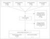
The search yielded 1365 abstracts. Two authors (AI and VG) selected those featuring, 1) medical students who were, 2) trained/educated using a, 3) genuinely hand-held device (as opposed to cart- or laptop-based ultrasound). We included primary and secondary research literature but not conference abstracts. References in relevant papers were searched manually to identify additional studies of interest. We identified 26 papers that fulfilled the inclusion criteria. Analysis of full texts led to the removal of 14 of these studies, which included 5 abstracts for which full text was not available. A flow diagram for the search is displayed in Fig. 1.
Data extraction and analysis
The following items were extracted from the papers selected for analysis: type of HHU device used; intensity, type, and duration of HHU-specific training offered; setting (clinical, e.g., ward-based or out-patient, or laboratory setting); pathologies detected/body areas scanned; metrics for the detection of left ventricular (LV) dysfunction; number of medical students involved and their year of medical studies, and any metrics reflecting student performance before and after the introduction of HHU-based scanning. We extracted sensitivity and specificity parameters, and calculated a diagnostic odds ratio (OR) for the detection of LV dysfunction using the HHU device. Data was collated and analysed using MetaDisc, a freely available statistical package designed for the meta-analysis of diagnostic data. The results were represented as forest plots and an analysis of heterogeneity was performed.
Results
Studies included
We retained nine primary literature articles for which full text articles were available.14)15)16)17)18)19)20)21)22) Three relevant non-systematic reviews were also identified,23)24)25) containing information on HHU use in undergraduate medical education, yielding a total of 12 studies (Fig. 1).
Studies were published between 2010 and 2014, three from USA14)15)16) and six from Europe.17)18)19)20)21)22) The mean number of medical student participants [standard deviation (SD)], based on studies where this information was available, was 18 (15.5), (range 1–45). The one study where the number of participants was unavailable, all students across four years of medical school participated.15)
Trainees
While all studies included medical students, 2 also included other healthcare professionals (medical residents22) and pharmacy residents16)), and did not separate the findings by type of participant in their final analyses. However, none of the participants had previous experience in ultrasonography thus qualifying as genuine “novices.”
Setting
Most studies were carried out on unselected patients in the hospital.16)17)18)20)21)22) Two studies were primarily classroom based, utilising volunteers14)19) with some inpatient scanning.14) The mean number (SD) of patients scanned, based on 6 studies for which the information was available, was 96 (65), range 27–211.
HHU devices used
Studies reported using 2 types of HHU device: Vscan (GE Vingmed Ultrasound AS, Horten, Norway) used in 6 studies, and Acuson P10 (Siemens Ultrasound, Surrey, UK) used in 3 studies. One of the studies did not report the model of HHU used.15) In all studies, results obtained by HHU examinations were compared to formal, comprehensive ultrasound examinations performed by an expert using high-end scanners in the local ultrasound department. We also noted that some literature featured hand-carried devices (such as the OptiGo and Micro-Maxx) and, incorrectly in our view, referred to them as handheld.25)
Training to use HHU
All studies reported hands-on, skills-teaching sessions, complemented by either didactic teaching (7 studies) or self-directed learning resources (1 study), or both (3 studies). One study used purely self-directed learning groups of students,14) while others utilised radiologists,18) sonographers,14) cardiologists and senior cardiology trainees,18) and even senior medical students, as teachers.15) When self-directed echocardiography simulators were used to teach image interpretation efficacy was lower than for a traditional lecture-based approach.14) The teaching itself varied in length and the mean (SD) duration of training was 9.8 (7.5) hours, range from 1 hour16) to 25 hours.21) There was no correlation between length of training and diagnostic accuracy assessed at the end of training, but such an association would have been difficult to detect due to the heterogeneity of the data and small sample size (Supplementary Fig. 1A, B, and C).
Fox et al.15) used on-line self-directed teaching materials and Apple iTunes-based podcasts to support learning. Another study compared the efficacy of traditional lecture-based approaches with fully self-directed online e-modules and self-directed simulation for the purposes of ultrasound education; no difference was detected with respect to understandings of theoretical aspects of ultrasonography and image interpretation.14) However, the presence of facilitators to guide the students resulted in better image acquisition abilities.
Clinical applications
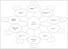
Medical students were taught to obtain and interpret ultrasound images of a variety of anatomical structures and organs (Fig. 2); others were trained to carry out the focused assessment with sonography for trauma (FAST) scan.19) The most widely studied application was imaging of the heart, featuring in 8 of the 9 primary literature articles. Medical students reported feeling confident to use the HHU device even after short (5 hours) training sessions.19)
A medical student trained to use a HHU device can achieve superior diagnostic accuracy compared with physical examination.17) Moreover, after only short training sessions (< 1 hr), novices can be guided ‘in real time’ to perform a focused cardiac examination under off-site expert guidance, with image quality comparable to that achieved by experienced sonographers.16) However, HHU devices achieve lower diagnostic accuracy than larger, more sophisticated scanners.21) Furthermore, medical students prefer cart-based devices to HHU and are able to obtain better quality images using the larger machines.19)
Effects of training
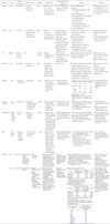
Assessment of the short-term effects of training varies between studies, making head-to-head comparison of results difficult. The most consistently detected pathology across the studies was LV systolic dysfunction. Sensitivity and specificity for the detection of LV dysfunction from four studies is summarised in the Supplementary Fig. 1A, B, and C. The summary receiver operating characteristic curve (Supplementary Fig. 2) demonstrates high diagnostic accuracy for detection of LV dysfunction. Although pooled sensitivity and specificity [95% confidence interval (CI)] for LV dysfunction reached 0.88 (0.83–0.92) and 0.86 (0.81–0.90) respectively, the data was heterogeneous (I2 values of 85.0% and 90.5% respectively). The diagnostic OR (95% CI) for the detection of LV dysfunction was 62.3 (12.8–303.8).
The sensitivity for the detection of pericardial effusions was variable, with one study reporting 100%,22) while the other reporting 40%.17) Similarly sensitivities for valvular regurgitation and stenosis varied (Table 1), but were consistently > 70%,17)22) aside from aortic regurgitation where one report demonstrates sensitivity to be 43%.17) Aortic root dilatation detection had the lowest sensitivity at 25% with a specificity of 88%.17) Diagnostic accuracy reached 100% for measurement of the abdominal aorta or for detection of ascites, and was at its lowest for detecting gallstones or cholecystitis.18)
To further emphasise the heterogeneity, some students were assessed using an objective structured clinical examination (OSCE) in a controlled environment, using written cases and a normal volunteer examination,19) while others were assessed based on a log of selected examinations, performed on non-standardised (authentic) patients on hospital wards.18) All studies confirmed that using HHU devices produced an improvement of the performance parameters monitored. The results are summarised in Table 1.
Discussion
Research into the use of HHU in medical education is scarce and highly heterogeneous. Studies focus on teaching medical students to identify pathology, mostly pertaining to the heart, rather than teaching anatomy and physiology. Nevertheless, we have found that teaching medical students the basics of ultrasound using novel HHU devices is feasible and effective. Many investigators recognise the use of self-directed learning packages to aid ultrasound learning and recognition of pathology. HHU is ‘the stethoscope of the future’ and may have multiple uses in the hands of the physician.2)26)27) While formal scanning with ‘full-blown’ ultrasound machines11)12) has been shown to increase the understanding of anatomy and physiology in undergraduate medical education, whether HHU has a similar effects has not yet been evaluated.
Benefits of HHU training for medical students and its effects
HHU is not a substitute for clinical examination, but rather an extension of it, improving diagnostic accuracy for a range of conditions.3)17)28) Although, data are inconsistent, high sensitivities are achieved for a number of cardiac and non-cardiac pathologies, which may not be detected on physical examination alone. Even with limited training, medical students across the studies demonstrated relatively high sensitivities (88%) and specificities (86%) for the detection of LV systolic dysfunction. It is fairly certain that HHU is always discriminatory between those with and those without LV systolic dysfunction, according to the odd's ratio (62.3).
Students also perform well when asked to complete a FAST scan and are able to detect free fluid on simulators as part of an OSCE, despite receiving only 5 hours of training.19) Abdominal aortic diameters20) can be measured accurately by novices, while the accuracy for the detection of pericardial effusions remains uncertain.17)22)
HHU can change patient management in emergencies when used by an expert.5) It is unlikely that a novice would be able to achieve the same accuracy. Nevertheless, diagnostic values are high when HHU is used by the students and may help with early recognition and management of the acutely ill patient (e.g., differentiating between LV dysfunction vs. acute exacerbation of chronic obstructive pulmonary disease.26) Familiarity with echocardiography during undergraduate medical education may also allow ‘tomorrow's doctors’ later to prioritise particular patients for standard ‘departmental’ echocardiogram, to recognise the limitations of the technique and, in some, to nurture a desire to further develop their skills in non-invasive imaging; all of these would support inclusion of HHU in the curriculum of medical schools.
Barriers to the widespread adoption of HHU in undergraduate medical education
Poor quality of available data to guide policy
There is substantial variation within the literature for all the parameters we extracted. There is no consensus on what defines the appropriate use of HHU in medical education. Until a position statement from relevant professional bodies is adopted, it is likely that penetration of HHU will remain patchy and inconsistent- driven mainly by local interest and resource.
Lack of consensus on the desirable level of competence
There is no generally held agreement about a minimal level of competence or a standard set of skills that should be taught. It may be possible to certify medical students in focused ultrasound scanning, but this approach has not been reported. Criteria and pathways for accreditation need to be developed and monitored to ensure appropriate clinical governance. Formal accreditation early in the career, with the provision of evidence of continuing use29) may be effective in maintaining competence in the longer term.
Limited availability of qualified teachers/trainers
Cardiologists and accredited cardiac sonographers are the ‘gold standard’ for teaching HHU of the heart, and are not easily released from their clinical duties to provide support for medical student education. Alternatives may be considered, such as non-clinical instructors, specially-trained students with an interest in imaging,15) junior doctors, and this may represent a fertile area for research and development. The most important factor for the success of integration of HHU teaching into the curriculum is a good team to drive the change.30) Free on-line HHU learning resources, such as podcasts,15) i-books,31) and e-modules,14) may have an important part to play.
Uncertainty about long-term retention of skills
Any attempt at disseminating ultrasound education using HHU in the medical school needs to be synchronised with a similar drive in the postgraduate arena. A recent study by Kimura et al.32) conducted on physician graduates shows that cardiac ultrasound skills decline within 2 years of non-use. Therefore, unless doctors have opportunities to maintain the ability to use HHU, it can be argued that undergraduate training is not worth the trouble.
Limitations
This review was limited to studies published in English. The studies were markedly inhomogeneous, which prevented rigorous comparisons. Most students included in the studies were volunteers, presumably with a specific interest in the topic, so results cannot be generalised to a situation where HHU would be ‘rolled-out’ to all students in a cohort.
Conclusion
It is possible to teach medical students how to use HHU scanners, and this enhances their diagnostic accuracy, especially for cardiac conditions. Current data on integrating HHU within medical curricula is suboptimal and highly heterogeneous. Further study is required to assess the longevity of skills retention for HHU, their precise role in the curriculum and in the development of medical careers, the financial impact of a HHU-based approach to medical curricula, pathways to accreditation, and to inform the development of consensus among educators and clinical leaders concerning the use of HHU.
 XML Download