This article has been
cited by other articles in ScienceCentral.
Keywords: Unroofed coronary sinus, Atrial septal defect
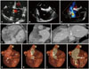
An 84-year-old woman with hypertension presented to our medical center with dyspnea and lower extremity edema. Electrocardiography demonstrated atrial fibrillation with a rapid ventricular response. Transthoracic echocardiography color Doppler showed abnormal flow in the region of the interatrial septum (
Fig. 1A). Transesophageal echocardiography demonstrated a defect adjacent to the interatrial septum (
Fig. 1B) with left-to-right flow (
Fig. 1C). Follow-up gated, 320-multidetector contrast-enhanced cardiac CT showed an isolated unroofed coronary sinus (
Fig. 1D, E, and F). Three-dimensional volume-rendered cardiac CT sequential cut planes further depicted a dilated, unroofed coronary sinus (
Fig. 1G-J).
To the best of our knowledge we report multimodality imaging findings in the oldest patient diagnosed with an unroofed coronary sinus atrial septal defect, the rarest atrial septal defect (< 1%) which accounts for 0.1% of all congenital heart diseases.
1) Of the four morphological types of unroofed coronary sinus this case illustrates type IV, partially unroofed terminal portion.
2) The multimodal approach of flow visualization with color Doppler echocardiography and anatomic assessment with 320-multidetector cardiac CT provided complementary imaging that facilitated evaluation of a rare congenital heart disease in a geriatric patient.
Figures and Tables
Fig. 1
Transthoracic echocardiography 4-chamber view color Doppler demonstrates abnormal flow along the interatrial septum (A, arrow). Transesophageal echocardiogram bicaval view shows a defect (arrow) adjacent to the interatrial septum in the expected location of the inferior vena cava (B) with color Doppler evidence of left-to-right flow (C). Contrast-enhanced 320-multidector cardiac CT inferior 4-chamber reformat shows contrast-opacified blood in the dilated coronary sinus draining into the right atrium (D). A biatrial reformat illustrates that the defect noted on the transesophageal echocardiogram (B, arrow) represents an unroofed terminal coronary sinus (E). The 2-chamber view shows a dilated, unroofed coronary sinus and a dilated left atrium. Sequential cut planes of volume-rendered images of the posterior heart depict the unroofed coronary sinus in 3-dimensions (F, arrow). The arrow points to a dilated coronary sinus (G, arrow). Sequential cut planes show an intact roof proximally (H, arrow) but an unroofed terminal coronary sinus (I, arrow). The unroofed coronary sinus drains into the right atrium (J, arrow). CS: coronary sinus, IAS: interatrial septum, LA: left atrium, LV: left ventricle, RA: right atrium, RV: right ventricle, SVC: superior vena cava.


 PDF
PDF ePub
ePub Citation
 Print
Print


 XML Download