

 PDF
PDF ePub
ePub Citation
 Print
Print


During the embryonic development of the heart, the two ventricles develop as independent structures on the two sides of the primitive plate. Normally, the two ventricles merge together at the apex and their cavities are separated by a bridge formed by muscle fibers (interventricular septum). An apical cardiac notch indicates the developing interventricular septum, and normally disappears later during organogenesis.1) Bifid cardiac apex arises when this process is abnormal, resulting in a defective union of the two ventricles at the apex and persistence of the cardiac notch. Bifid cardiac apex is a common finding in sea mammals like whales and manatees, however this abnormality is very rare in humans.1)2) Only few cases have been reported in the literature, most of which were accompanied by additional congenital cardiac abnormalities.1)2)3)4)
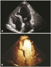
We are presenting the images of a 73-year-old woman with bifid cardiac apex who was admitted in our Department with palpitations, dizziness and diaphoresis. Her past medical history included a transient ischemic attack 5 years ago. Cardiac examination revealed regular rate and rhythm with normal heart sounds, without any murmurs. The systemic physical examination was unremarkable. Chest X-ray was clear with a normal cardiac shillouette. ECG revealed minimal ST-T depression on precordial leads. Ambulatory ECG showed an episode of slow ventricular tachycardia at 105 bpm with a duration of 20 seconds and four shorter ventricular runs. Transthoracic 2D and contrast echocardiography (Fig. 1, Supplementary movie 1, 2, and 3) demonstrated a cleft-like small chamber between the left and right ventricles (bifid left ventricle) with normal systolic function, similar thickness to normal myocardium, and normal perfusion. The right ventricle seemed hypoplastic with adequate systolic function. The atria were of normal size. No additional heart abnormality was detected with echocardiography. Considering the above findings, the lack of a history of coronary artery disease or myocardial infarction, and the absence of Q waves on ECG, we feel that our finding represents true bifid left ventricle/bifid cardiac apex, and that the possibility of ventricular pseudoaneurysm or aneurysm due to prior myocardial infarct related to coronary artery disease is extremely unlikely. Further investigation with a cardiac MRI was suggested, but the patient refused any further tests and was discharged on antiarrhythmic and anticoagulant therapy.
Bifid cardiac apex is a very rare developmental abnormality of the heart in humans, and its clinical significance and impact on survival is currently unknown. There has been some controversy whether this abnormality is a potential source of embolism or arrhythmias. Very few patients with bifid cardiac apex have been reported in the literature so far. In a case series of 3 patients, two patients died shortly after birth and one during early adulthood, but all 3 patients had additional severe and multiple congenital heart defects.1) A bifid apex was confirmed with autopsy in a 11-year-old boy who died suddenly while he was playing, presumably due to malignant arrhythmia.4) However, this boy also had an aberrant right coronary artery with high take-off from the ascending aorta and slit-like orifice, and therefore the role of the bifid cardiac apex for his sudden cardiac death was unclear.4) Two additional cases of bifid apex have been reported in young patients with additional congenital heart defects, without any data regarding their follow-up.2)3) Interestingly, a bifid cardiac apex without any additional congenital heart defect has been confirmed with autopsy in a young adult who died from combined drug toxicity.5)
Our image illustrates a rare case of a bifid cardiac apex in an elderly woman without significant symptoms, and implies that this rare developmental abnormality of the heart may not be very malignant when no additional congenital cardiac abnormalities are present.
 XML Download