

 PDF
PDF ePub
ePub Citation
 Print
Print


Introduction
Degenerative mitral regurgitation (MR) is the most common form of organic MR disease in developed countries.1) Surgical repair of the mitral valve (MV) remains the best treatment option for patients with severe degenerative MR,2) which is associated with good long-term follow-up.3) However, degenerative MR disease still carries a sizable risk of recurrent MR after surgery. This seems to be related to the progression of left ventricular (LV) and mitral annular (MA) disease.4) Both left atrial (LA) enlargement and remodeling are compensatory mechanisms in patients with severe MR. In patients with severe MR, LA size is an important predictor of outcome in both conservative treatment and after MV surgery.5)6) Moreover, one of the predictors of successful MV repair is the extent of MA disease.7) A previous study using a Laplace's law model indicated that curvature of the leaflets is a beneficial feature. Thus, saddle shape preservation decreases leaflet and annular strain and also increases leaflet coaptation.8)9) In another study, a late systolic decrease in MA non-planarity can additionally increase leaflet stress in patients with MR, resulting in elongation and secondary chordae rupture.9) Consequently, the extent of MA remodeling and dysfunction is thought to be correlated with MR severity. MA remodeling and contractile dysfunction are also associated with LA dilation and LV dilation. Generally, a decrease in LA size, or reverse remodeling, has been observed after MV surgery. LA reverse remodeling occur after surgery of MR with preserved LV ejection fraction (LVEF).10) One previous study concluded that MA dysfunction might be a prognostic factor for increased repair durability and MR recurrence after successful MV repair.11) Therefore, thorough assessment of MA geometry and function as well as of the determinants of MA remodeling are pivotal to understanding the pathophysiology and severity of MR, and to planning effective reparative surgery.
Two-dimensional echocardiography (2DE) guides the timing of surgical intervention by providing information on MR severity, monitoring LV and LA remodeling, and estimating pulmonary artery systolic pressure.12) Currently, the only recommendation for the use of echocardiography in MA assessment before surgery is for the measurement of its anteroposterior diameter on LV 2DE long axis view.13) In addition, MA quantitative assessment that completely changed our way of assessing MA shape and function by the advent of 3-dimensional echocardiography has been used for a better understanding of MR pathophysiology, 14) and on following up patients.15) However, the dynamics of the MA in MR remains controversial.16) The relationship between the function of the MA and MR severity has not been reported, and the connection between MA remodeling and left heart chamber size and function remains to be defined. The aim of this study was to identify additive prognostic factors that could be used for timing surgical assessment of MA non-planarity using real-time 3D transesophageal echocardiography (RT3D-TEE) in patients with severe chronic primary MR and preserved LV systolic function.
Methods
Patient selection
Forty-seven patients with severe chronic primary MR and preserved LVEF (> 60%) scheduled for MV repair were enrolled between March 2010 and March 2012. Serial 2D TTEs were performed before surgery, 2 weeks after surgery and at least 6 months postoperatively. RT3D-TEE was also performed just before operation and in the operating room immediately post-operative. The severity of MR was determined via integrated echocardiography evaluation using the following measurements: LV dimension, effective regurgitant orifice (ERO) and regurgitant volume. Severe MR was defined as an ERO > 40 mm or a regurgitant volume > 60 mL.17)18) Exclusion criteria included secondary MR due to either a distortion of the subvalvular apparatus or LV enlargement and remodeling (idiopathic cardiomyopathy or ischemic heart disease); other concomitant valvular disease of moderate or severe severity; coronary artery disease (defined as > 50% narrowing in at least one coronary artery in a previous angiogram); history of myocardial infarction; coronary artery bypass graft; acute coronary syndrome; atrial fibrillation; chronic renal failure or suboptimal imaging.
The regional ethics committee approved the study. The patients provided informed consent prior to enrollment.
Two-dimensional transthoracic echocardiography
2DE was performed using commercially available equipment (Vivid 9; GE Medical Systems, Milwaukee, WI, USA). End diastole was defined as the frame with the largest cavity area immediately before the onset of QRS, while end systole was the frame with the smallest cavity area. The LV end-diastolic dimension (LVEDD), LV end-systolic dimension (LVESD), diastolic interventricular septum thickness and diastolic LV posterior wall thickness were all obtained from the parasternal views, according to standard guidelines.19) LV mass was calculated from the linear dimensions using the American Society of Echocardiography recommended formula. LA volume was measured at end-systole from the frame just preceding MV opening using the biplane area length method in the apical 4- and 2-chamber views, and was indexed to body surface area (BSA).19) LV diastolic function was assessed by the early (E wave) and late (A wave) transmitral velocities, the corresponding E/A ratio, and the E wave deceleration time using pulsed-wave Doppler. Tissue Doppler imaging was used to measure peak early diastolic MA velocity (e′) at the septal mitral annulus in the apical 4-chamber view. An E/e′ ratio was calculated to noninvasively estimate LV filling pressure.20) We used the average of three consecutive Doppler signals to take these measurements.
Quantitative and qualitative measures of MR severity were taken according to the American Society of Echocardiography guidelines.18) MR regurgitant volume was calculated using the proximal isovelocity surface area (PISA) method. The ERO area was determined by dividing the regurgitant flow rate (calculated as 2 πr2 × aliasing velocity, where r is the PISA radius) by the peak MR velocity.12)
Real-time three-dimensional transesophageal echocardiography
The RT3D-TEE images of the MV were obtained in full-volume mode, in which electrocardiographically triggered wedge-shaped subvolumes were obtained over seven consecutive cardiac cycles using an iE33 system equipped with a matrix probe (X7-2t; Philips Medical Systems, Andover, MA, USA). And, the RT3D-TEE images of patients with atrial fibrillation were obtained during a single cardiac cycle.
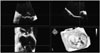
Full-volume 3D data sets were digitally stored and transferred to a workstation with Q-Laboratory Mitral-Valve-Quantification Software (Philips Medical Systems, Bothell, WA, USA) for offline analysis. Three orthogonal mitral annulus images were displayed and subsequently modified to optimize visualization of the entire annulus (Fig. 1). MA measurements were performed six times during the cardiac cycle in early, middle, and late diastole and early, middle, and late systole. Measurements of annular diameter, annular perimeter, annular area and annular height were performed during late systole. Early diastole was identified with MV opening, late diastole before mitral closure, and middle diastole as midway between these frames. Early systole was identified just before mitral closure, late systole on the frame preceding aortic closure, and middle systole midway between these frames.
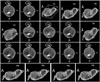
The resulting 3D representation was adjusted to visually match the anatomy viewed in 3D and 2D cut-plane views. From this model, several parameters were calculated (Fig. 2): 1) annular geometry: annular area, as the area of the minimal surface spanning the annulus and the anteroposterior, anterolateral and posteromedial diameters; annular height, as the distance along the atrial-ventricular direction between the lowest and highest point of the annulus; and the planarity index, defined as the ratio of the height to anterolateral-posteromedial diameter, equal to zero for a flat MV and increasing when the MV's saddle shape was more pronounced; 2) leaflet size: exposed 3D area of the anterior or posterior leaflet, as well as the 3D total exposed leaflet area, as the sum of the two previous measurements; 3) coaptation geometry: the length of the coaptation line projected to approximate the leaflet surface, the area of the region where the anterior and posterior leaflets overlap and the mean height of the same region; and 4) the aortic-to-mitral plane angle.
Measurement variability was determined by repeating the measurements on stored 3D data sets at least 1 week after the initial measurements by the same observer (intraobserver) and a different observer (interobserver).
The average value of baseline annulus height/BSA was 4. And, we defined definitions of decreased annular height/BSA to be less than 4.
Follow-up
Data were obtained until August 2012 (mean follow-up duration: 5.43 months) within 2 weeks after regular outpatient visits and at least 6 months postoperatively. Particular care was taken to obtain information regarding the development of symptoms, eventual MV repair or replacement, and deterioration of LV function.
Statistical analyses
Continuous variables are listed as mean values. Categorical variables are presented as frequencies and group percentages. Continuous variables were compared using the Student's t-test. The chi-square or Fisher's exact test was used for comparison of categorical variables. A 2-tailed p value < 0.05 was considered statistically significant. Pearson's correlation coefficient (r) and intraclass correlation coefficient (ICC) were calculated to express agreement between LA volume index (LAVI) and the annular height/BSA obtained using RT3D-TEE. Interobserver agreement was demonstrated by calculating the coefficient of variation of repeated measurements and the ICC. All p values < 0.05 were considered significant. Data analysis was performed utilizing SPSS version 18.0 (SPSS Inc., Chicago, IL, USA).
Results
Pre-LAVI, delta LAVI (pre-post) and annular height/BSA on RT3D-TEE correlation
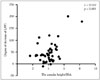
Mean patient age was 55.4 ± 15.1 years and 24 (51.1%) were male. The annular height/BSA obtained from RT3D-TEE was correlated with delta LAVI (pre-post) [r = 0.519, p < 0.001, 95% confidence interval (CI) 0.273–0.702] (Fig. 3).
Baseline characteristics and echocardiography in severe MR with decreased or normal annular height/BSA
The study subjects were then divided into 2 groups based on average annular height/BSA: normal annular height/BSA and decreased annular height/BSA. Baseline characteristics and echocardiographic parameters are shown in Table 1 and 2. None of the baseline characteristics except for sex and BSA were significantly different when comparing the normal and decreased annular height/BSA groups. The decreased annular height/BSA group had a higher number of men than normal annular height/BSA group (p = 0.017). Additionally, the decreased annular height/BSA group had a higher BSA than the normal annular height/BSA group (p = 0.003). Echocardiography parameters of the normal and decreased annular height/BSA groups were also compared and were not significantly different between the two groups.
Analysis of postoperative echocardiography
After at least 6 months of postoperative follow-up, patients with decreased annular height/BSA had larger LVEDDs and LVESDs than patients with normal annular height/BSA (p = 0.002 and 0.038). Patients in the decreased annular height/BSA group also had higher LAVIs than patients in the normal annular height/BSA group (p = 0.047).
In addition, patients with decreased annular height/BSA had larger pre-post2 LVEDDs and pre-post2 LAVIs than patients with normal annular height/BSA (p = 0.045 and 0.040) (Table 3).
Real-time 3D transesophageal echocardiography analysis
None of the preoperative baseline characteristics except for annular height to commissural width ratio (AHCWR) were significantly different when the normal and decreased annular height/BSA groups were compared. The decreased annular height/BSA group had a smaller AHCWR than the normal annular height/BSA group (p < 0.001) (Table 4).
Evaluation of the postoperative RT3D-TEE results revealed that none of the parameters aside from the AHCWR were significantly different in the normal and decreased annular height/BSA groups. The decreased annular height/BSA group had a smaller AHCWR than the normal annular height/BSA group (p = 0.026) (Table 5).
Independent predictors of postoperative LA remodeling in patients with severe MR
Univariate linear analysis revealed that the preoperative annular height/BSA (95% CI, 1.167–8.009; p = 0.023) independently predicted postoperative LA remodeling (postoperative LAVI reduction of less than 34 mL/m2). Furthermore, postoperative LVEDD (95% CI, 1.020–1.458; p = 0.029) was also independently predictive of postoperative LA remodeling (postoperative LAVI reduction of less than 34 mL/m2).
In multivariable analysis, preoperative annular height/BSA (95% CI, 0.005–0.768; p = 0.030) independently predicted postoperative LA remodeling (postoperative LAVI reduction of less than 34 mL/m2) (Table 6). To summarize, we found that LA reverse remodeling occurs frequently after surgery for severe MR with preserved LVEF, and that preoperative annulus height is the only variable significantly associated with a postoperative LAVI reduction of less than 34 mL/m2.
Discussion
The main findings of our study of patients with severe degenerative MR and preserved LVEF were: 1) after at least 6 months of postoperative follow-up, patients with the decreased annular height/BSA had larger LVEDDs and LVESDs than patients with normal annular height/BSA. Patients with decreased annular height/BSA had larger LAVIs, pre-post LVEDDs, and pre-post LAVIs than those with normal annular height/BSA. 2) The decreased annular height/BSA group had a lower AHCWR than the normal annular height/BSA group. 3) LA reverse remodeling occurs after surgery for severe degenerative MR with preserved LVEF, and preoperative annulus height/BSA was the only variable significantly associated with postoperative LAVI reduction less than 34 mL/m2.
LAVI changes and postoperative LA remodeling or dysfunction in patients with severe degenerative MR and preserved LVEF
Both LA enlargement and remodeling are compensatory mechanisms in patients with severe MR. In patients with severe MR, LA size seems to be an important predictor of outcomes after conservative treatment or MV surgery.5)6)21) In a previous study, Grewal et al.22) showed that the patients with MR present a loss of early MA contraction, despite the same magnitude of LV contraction, and suggested ventriculo-annular decoupling. Another study suggested that MA size and reduced fractional area shortening were more related to LA rather than to LV size and dysfunction, at least in cases of degenerative severe MR with preserved LVEF.22) A decrease in LA size, or reverse remodeling, has been observed after MV surgery.10) In the present study, the LAVI obtained using echocardiography at least 6 months postoperatively was smaller in the normal annular height/BSA group than in the decreased annular height/BSA group. Moreover, the degree of LAVI decrease between baseline and at least 6 months postoperatively was greater in the normal annular height/BSA group. Thus, annular height/BSA might be an additive predictive factor for postoperative LA dysfunction or remodeling. In this way, assessment of MA geometry and function and the determinants of MA remodeling in MR have become pivotal to understanding the pathophysiology of severe MR and to planning effective reparative surgery.
Early detection of postoperative LA remodeling or dysfunction in patients with severe degenerative MR and preserved LVEF
Surgical intervention for severe MR is usually triggered by the occurrence of symptoms, declining LV function, significant LV enlargement, or by the development of atrial fibrillation or severe pulmonary hypertension.23) The extent of MA remodeling and dysfunction has recently been correlated with the severity of MR. A previous study found lower MA displacement in patients with MR than in normal patients.16) Unique annular shape is considered to be important in reducing leaflet stress and enhancing valve competence during systole.24)
A previous study using Laplace's law model indicated that curvature of the leaflets is a beneficial feature. Saddle shape preservation decreases leaflet and annular strain while increasing leaflet coaptation.8)9) In another study, MA non-planarity was reported to have a role in reducing MV leaflet stress.24) Late systolic decrease in MA non-planarity can additionally increase leaflet stress in patients with MR, causing elongation and secondary chordae rupture.9) As expected, MV tenting height and volume decreased from mid-to end-systole in patients with MR due to progressive mitral leaflet prolapse. This also supports the hypothesis that MA non-planarity further reduces peak leaflet stress.25)
To summarize, annular flattening and enlargement has been shown to decrease leaflet curvature, resulting in increased leaflet stress and strain, which over time may cause MV degeneration, chordal rupture, and MR.11)
The mitral annulus of patients with severe degenerative MR is not only greater in area, but also flatter, with a decreased AHCWR. In a previous study, this ratio was shown to be strongly associated with chordal rupture, the prevalence of which progressively increased from 7% in those with the most saddle-shaped annuli to 42% in those with the most planar. Annular flattening is also associated with increased leaflet billowing volume. Taken together, these variables determine MR severity.25)
In previous studies, when used to quantify the changes in MA non-planarity in chronic MR, the AHCWR and the non-planarity angle have shown a similar inverse relation. And, the AHCWR showed a favorable correlation with the non-planarity angle. Contrariwise, an increase in the saddle configuration of the mitral annulus is demonstrated as an increase in the AHCWR. The geometric description of the non-planarity angle made it intuitive that changes in the AHCWR will be associated with an inverse change of the non-planarity angle. And an increase in the annular height was associated with a decrease of the non-planarity angle.26)
MA non-planarity has been already reported to have a role in reducing MV leaflet stress. So, the previous study also revealed that annulus flattening and contractile dysfunction progressed in parallel with severe enlargement of the LA and MA.27)
Previous studies showed that RT3D-TEE was both more accurate and more reproducible than 2D imaging for the assessment of MV lesions. In our study, the decreased annular height/BSA group had a smaller annular height and AHCWR than the normal annular height/BSA group in baseline and 6-month postoperative RT3D-TEE images.
Additionally, a prior study showed that RT3D-TEE provided incremental diagnostic value, particularly in the assessment of complex multisegmental MV disease involving one or both leaflets compared to simple monoleaflet lesions.28) Therefore, RT3D-TEE is a comprehensively balanced technique for describing functional MV anatomy and may be provided additional information predictive of postoperative LA remodeling and postoperative prognosis.
Limitations
Several potential limitations of our study must be noted. First, this was a single-center study that included a relatively selective population of patients with chronic primary severe MR without other concomitant valvular diseases. Consequently, the sample size was relatively small. Second, patients with severe MR and decreased annular height had larger BSA compared with those with normal annular height. However, BSA was not significantly associated with annular height variance. Third, only patients with chronic primary severe MR and preserved LV systolic function were enrolled. Therefore, our results cannot be extended to patients with chronic severe MR and LV dysfunction. In the future, large-scale prospective studies are needed to assess MA height in chronic primary severe MR with LV dysfunction. Finally, we did not investigate long-term follow-up echocardiographic data in the current study; only data collected at least 6 months postoperatively were analyzed. Ongoing study of this topic at our institution will include long-term follow-up data, with the intention of elucidating the prognostic role of MA height in the future.


 XML Download