Journal List > J Cardiovasc Ultrasound > v.24(4) > 1018455

Alkema, Spitzer, Soliman, and Loewe: Multimodality Imaging for Left Ventricular Hypertrophy Severity Grading: A Methodological Review

Abstract

Left ventricular hypertrophy (LVH), defined by an increase in left ventricular mass (LVM), is a common cardiac finding generally caused by an increase in pressure or volume load. Assessing severity of LVH is of great clinical value in terms of prognosis and treatment choices, as LVH severity grades correlate with the risk for presenting cardiovascular events. The three main cardiac parameters for the assessment of LVH are wall thickness, LVM, and LV geometry. Echocardiography, with large availability and low cost, is the technique of choice for their assessment. Consequently, reference values for LVH severity in clinical guidelines are based on this technique. However, cardiac magnetic resonance (CMR) and computed tomography (CT) are increasingly used in clinical practice, providing excellent image quality. Nevertheless, there is no extensive data to support reference values based on these techniques, while comparative studies between the three techniques show different results in wall thickness and LVM measurements. In this paper, we provide an overview of the different methodologies used to assess LVH severity with echocardiography, CMR and CT. We argue that establishing reference values per imaging modality, and possibly indexed to body surface area and classified per gender, ethnicity and age-group, might be essential for the correct classification of LVH severity.

Introduction

Left ventricular hypertrophy (LVH), defined by an increase in left ventricular mass (LVM), is a common cardiac disorder with an estimated echocardiographic prevalence of 36–41% in patients with hypertension, which increases with age, hypertension severity and obesity.1)2)3) LVH can be caused by an adaptation of the myocardium to increased pressure or volume load, genetic mutations or systemic conditions, as summarized in Table 1.3)4)5)6)7)8)9)10)11)12)13)14)15)16) LVH develops gradually, and therefore patients remain asymptomatic in early stages. When the condition progresses, symptoms related to diastolic and systolic dysfunction ultimately bring the patient to the clinic.6) Analysing the grade of LVH severity is of great importance to clinical practice in terms of prognosis and treatment choices. An increased LVM is strongly linked to an increased risk of cardiovascular events.2)17) The three main non-invasive cardiac imaging techniques used to assess the severity of LVH are echocardiography, cardiac magnetic resonance (CMR) and computed tomography (CT).18) With this manuscript, we provide a comprehensive methodological review of different techniques for the assessment of symmetrical LVH based on the following parameters: wall thickness, LVM and left ventricle (LV) geometry. Furthermore, we provide a simplified proposal for inter-technique standardization. We aim to summarize LVH reference values, indexed by body surface area (BSA) and classified per gender for optimal classification of LVH severity using different imaging modalities.

Pros and Cons of Imaging Modalities for Assessing LVH Severity

Echocardiography

Echocardiography was introduced in the 1950s and has become the cornerstone of non-invasive imaging of the heart. This currently widespread technique is based on ultrasound waves directed to the heart, which are reflected and translated into several modalities that can be used to assess LVH, namely: M-mode echocardiography; two-dimensional echocardiography (2DE), and three-dimensional echocardiography (3DE) images.19)20) The most important advantages of echocardiography are its wide accessibility, lack of radiation exposure, and excellent temporal resolution, which is superior to all other imaging techniques. M-mode echocardiography, often used for LVH assessment, has a temporal resolution up to 1 ms. However, echocardiography is operator and patient dependent, which could result in a relatively high inter-observer variability.20)21)22) Furthermore, geometric assumptions represent an important limitation in LVM estimation in M-mode and 2DE, but this can be overcome by the use of 3DE: when 3DE measurements of LVM are compared to CMR measurements as gold standard, they correlate better, and have smaller limits of agreements, than LVM measurements with 2DE.23)24) Although overestimation used to be a problem in 3DE LVM measurements, Shimada and Shiota25) showed that accuracy of 3DE in LVM measurement has significantly increased over time, which was recently confirmed by Mizukoshi et al.,26) who showed an excellent accuracy, correlation and agreement between 3DE and CMR LVM measurements.
In most patients, transthoracic echocardiography allows for an adequate assessment of the heart. Nevertheless, poor endocardial delineation due to insufficient image quality such as in patients with emphysema could be solved by using either transoesophageal approach or the use of contrast media.18)21)

Cardiac magnetic resonance

CMR became available in the beginning of the 1980's27) and uses the nuclear magnetic resonance of hydrogen to identify specific tissues.28) Characteristically, it is not dependent on acoustic windows, measurements are less operator dependent, and overall resolution is adequate.21)22) This technique is considered the gold standard for assessment of most cardiac parameters including LVM, also allowing for cardiac tissue characterization. Notwithstanding, due to its high costs and lower availability, it is far less utilized than echocardiography.21)22) Furthermore, CMR has a relatively low spatial resolution (compared with CT), prolonged examination time, and is relatively contraindicated in patients with mechanical devices (e.g., pacemakers, prosthetic valves).21)22)

Computed tomography

CT utilizes X-rays that are sent through the body and absorbed at different intensities depending on the tissue. Detectors on the other side of the body identify the remaining X-rays, which allows for tissue differentiation.29) While the first CT scanners in the 1970's had only one detector and provided consequently a rather limited field of view per rotation, modern CT scanners have at least 64 detectors, and in the most advanced machines this number can reach even more than 700.29) The shift from electrocardiographic (ECG)-gated retrospective acquisitions towards ECG-triggered prospective acquisitions for the purpose of radiation dose reduction, limits the availability of end-diastolic phases, as preferably mid-diastolic phases are captured.30) Until now, no data is available regarding reference values for parameters such as wall thickness in mid-diastole. Cardiac CT is predominantly used for excluding the presence of coronary artery disease in patients with intermediate risk.31) However, the use of cardiac CT is expected to expand rapidly, since it allows for anatomical and functional evaluation of coronary lesions, and facilitates decision making before an invasive procedure.32)33) Furthermore, CT is used when a patient has contra-indications for CMR, offering an excellent spatial resolution and unrestricted field of view. However, the relatively low temporal resolution and the radiation exposure (ranging from 5–20 mSv21)22)) make cardiac CT the least preferred technique among the three for LVH assessment.21) Table 2 summarizes the main technically and clinically relevant characteristics of echocardiography, CMR and CT.

Methodology of LVH Assessment within Different Techniques

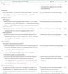
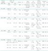
Guidelines with reference values for cardiac chamber quantification are available for echocardiography,18) but not yet for CT and CMR. Consequently, the reference values provided by these guidelines for echocardiography are used in the clinic for CMR and CT. The three main parameters for the assessment of LVH severity are wall thickness, LVM and LV geometry.18) Table 3 summarizes relevant parameters with cut-off values for LVH severity classification from the guidelines for echocardiography and available original reports for CMR and CT.18)34)35)36)37)

LV thickness

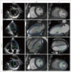
According to the guidelines for chamber quantification using echocardiography,18) LV posterior wall thickness is an important indicator for LVH severity. LV posterior wall, together with the LV internal diameter is used to calculate the relative wall thickness (RWT) as RWT = 2 × posterior wall thickness / LV internal diameter at end diastole. On the other hand, septal wall thickness gives an indication of the presence and severity of LVH.18) The latter parameter is often used in clinical practice.38) In echocardiography, LV thickness quantitation is performed at end-diastole (the frame before mitral valve closure or the frame in the cardiac cycle in which the ventricular dimension or volume is the largest). Standard LV thickness measurements could be performed using either M-mode or 2D linear diameters.18) The posterior wall is measured in the parasternal long-axis or short-axis view at or immediately below the level of the mitral valve leaflet tips, which is approximately at the junction of the basal- and mid-inferolateral segments (Fig. 1). Likewise, both views are used for the measurement of the septal wall, corresponding to the boundary between the basal- and mid-anteroseptal segments (Fig. 1).18) In routine practice, although not formally recommended, septal thickness is often measured in the 4-chamber view, at the junction between the basal and mid inferoseptum, or simply at the level of the thickest portion. The appropriateness of this 2D measurement would require further validation. For CMR, the same reconstruction protocols based on 2D non-contiguous coverage of the LV or 3D whole-heart imaging, recreate those end-diastolic views used for echocardiography.39) Thus, CMR linear measurements are obtained from the same segments as recommended for echocardiography. However, for CT, the views available for LVH assessment vary according to local reconstruction protocols. In principal, CT multiplanar reconstructions could reproduce the same segmentation used for echocardiography and CMR. Current software applications for CT- and CMR-based LV function and LVM assessment require contour delineation of the LV, and provide results of LV thickness using the 16-segment model, as well as LVM.40)

LVM

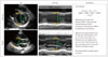
LVM assessment with all three techniques requires quantification of myocardial end-diastolic volume which is calculated either through geometric formulas (when derived from M-mode or 2D images) or directly measured (in 3D imaging) (Fig. 1 and 2). After the end-diastolic myocardial volume is calculated, it is converted to mass by multiplying it with the myocardial density (approximately 1.05 g/mL).18) To generate the LVM index (LVMi), the LVM is divided by BSA (resulting in the unit g/m2).

LV geometry

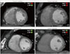
Determination of the LV geometry according to guidelines requires the LVMi and RWT as described above.6)18) With regards to geometry, the LV can be described as normal, concentric remodelled, and concentric or eccentric hypertrophied, as depicted in Fig. 3 and Table 3. Other classifications have been suggested, possibly increasing the prognostic value of LVH based on dilatation.41)42) As this review focuses on methodology of guideline based assessment of LVH severity, we would like to point interested readers to references on this important topic.41)42)

Inter-Technique Agreement and Disagreement

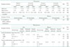
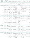
Although the methods for chamber quantitation may apply for all techniques, differences in spatial and temporal resolution can account for variations in reference values, as shown by comparative studies,37)43)44)45)46)47)48)49)50)51)52)53)54)55)56) of which the most important results are summarized in Table 4 and 5 for wall thickness and LVM, respectively. The comparative studies show that agreement is acceptable between techniques. When echocardiography is compared to CMR or CT in the measurement of wall thickness and LVM, generally a weaker Pearson's correlation and a larger bias with accompanying limits of agreement are found37)43)44)45)46)47)53)54)55) than when CMR and CT are compared,48)49)50)51)56) indicating that the most important drawback of echocardiography is the larger operator dependence, as compared to CMR and CT; most likely caused by the lower spatial resolution of echocardiography. Noteworthy, a difference of 2 mm in septal wall thickness could reclassify the presence and/or the severity of LVH (Table 3).6)18) Interestingly, although CMR is considered the gold standard for measurement of most cardiac parameters, CT is known to be the least operator dependent when it comes to measurements of for example the aorta and aortic annulus.57)58) The studies cited in Table 4 and 5, however, do not show a clear difference in operator dependence when CMR and CT are compared, indicating that for the parameters that are used for LVH severity assessment, both CMR and CT will suffice if the quality of echocardiographic measurements of parameters that are relevant for LVH severity assessment are not sufficient.

Gender, Ethnicity, Age, and Body Surface Area

Several studies have shown that gender, age, body size, and ethnicity influence reference values for cardiac parameters.2)18) Accordingly, reference values should be examined with caution. Indexing the LVM to BSA or heightx (where x stands for allometric powers such as 1.7, 2.13, or 2.7) allows for better comparison of persons with different body sizes. However, the optimal indexing method remains controversial. Most studies have used LVM/BSA, nevertheless recent data suggests that indexing LVM to heightx may more accurately predict events in obese patients.18) Paucity of data in this regard prompts the need for continued research in populations with normal and hypertrophied hearts.

Conclusion

LVH severity grading is important for diagnosis, prognosis and guidance of therapy. Three main parameters including wall thickness, left ventricular mass, and left ventricular geometry, are easily assessed with echocardiography, CT or CMR. Data representing normal populations is abundant for echocardiography, however still limited for CT or CMR. A standard methodology for LVH severity grading among modalities is advised to further improve comparability and facilitate clinical decision making.

Figures and Tables

Fig. 1

Methodology for the measurement of the septal and posterior wall thickness, LV mass, and LV diastolic diameter with echo, CMR and CT. Short axis: measurements of septal and posterior wall thickness (yellow lines). 2-chamber, 4-chamber, and short axis views: contour drawing of the LV for three-dimensional LVM measurement (blue lines). Contour drawing may primarily be performed in short axis images (all available) and confirmed with long axis views; or, using primarily long axis views while rotating the LV on its longitudinal axis, and confirmed with short axis views (all available). LV (end-)diastolic diameter (green lines). Echo: echocardiography, CT: computed tomography, CMR: cardiac magnetic resonance, LV: left ventricle, LVM: left ventricular mass.

jcu-24-257-g001
Fig. 2

Calculation of LV mass with an echocardiographic linear method. LV: left ventricle.

jcu-24-257-g002
Fig. 3

LV geometry patterns. RWT = 2 × posterior wall thickness / LV internal diameter at end diastole. LVMi = LVM / BSA. Normal = green, abnormal = red. Abnormal RWT is defined as > 0.42, abnormal LVMi is > 115 g/m2 (men), and > 95 g/m2 (women).18) RWT: relative wall thickness, LVMi: left ventricular mass index, LV: left ventricle, LVM: left ventricular mass, BSA: body surface area.

jcu-24-257-g003
Table 1

Overview of most important aetiologies of LVH

jcu-24-257-i001

LVH: left ventricular hypertrophy, BMI: body mass index, CMR: cardiac magnetic resonance, CT: computed tomography, LVM: left ventricular mass, LV: left ventricle

Table 2

Comparison of imaging modalities used for LVH severity grading

jcu-24-257-i002

LVH: left ventricular hypertrophy, echo: echocardiography, CMR: cardiac magnetic resonance, CT: computed tomography

Table 3

LV geometry and wall thickness cut-off values in guidelines and original studies for different imaging techniques

jcu-24-257-i003

*RWT = (2 × posterior wall thickness / LV internal diameter at end diastole). LVMi = [(myocardial end-diastolic volume × myocardial density) / body surface area], RWT = [(inferolateral + septal wall thickness at end-diastole) / LV end-diastolic diameter]. LVMi = as guidelines. Increased RWT was defined as > 0.45 mm, increased LVMi as > 81 g/m2 (men) or > 62 g/m2 (women), LVMi: as guidelines. Criteria for abnormal LVM: above the 97th percentile for LVMi. 2DE: two-dimensional echocardiography, CMR: cardiac magnetic resonance, CT: computed tomography, GL: guidelines, LV: left ventricle, LVMi: left ventricle mass index, OR: original research, RWT: relative wall thickness (mm), SD: standard deviation, UL: upper limit, -: not available

Table 4

Non-invasive imaging techniques compared in assessment of wall thickness

jcu-24-257-i004

2/3DE: two/three-dimensional echocardiography, BA: Bland-Altman analysis, BAL: basal anterolateral, CC: correlation coefficient, cm: centimetre, CT: computed tomography, CCTA: cardiac CT angiography, CI: confidence interval, CMR: cardiac magnetic resonance, ED: end-diastole, ES: end-systole, HCM: hypertrophic cardiomyopathy, LV: left ventricle, LOA: limits of agreement, Post.: posterior, r: Pearson correlation coefficient, SD: standard deviation, Segm.: segmental, Sept.: septal, WT: wall thickness, -: not available

Table 5

Non-invasive imaging techniques compared in assessment of left ventricular mass

jcu-24-257-i005

2/3DE: two/three-dimensional echocardiography, BA: Bland-Altman analysis, CC: correlation coefficient, CT: computed tomography, CCTA: cardiac CT angiography, CI: confidence interval, CMR: cardiac magnetic resonance, HCM: hypertrophic cardiomyopathy, LOA: limits of agreement, LV: left ventricle, LVH: LV hypertrophy, LVM: left ventricular mass, LVMi: left ventricular index, MDE: myocardial delayed enhancement, r: Pearson correlation coefficient, SD: standard deviation, -: not available, EF: ejection fraction

References

1. Cuspidi C, Sala C, Negri F, Mancia G, Morganti A. Italian Society of Hypertension. Prevalence of left-ventricular hypertrophy in hypertension: an updated review of echocardiographic studies. J Hum Hypertens. 2012; 26:343–349.
2. Ruilope LM, Schmieder RE. Left ventricular hypertrophy and clinical outcomes in hypertensive patients. Am J Hypertens. 2008; 21:500–508.
3. Cuspidi C, Rescaldani M, Sala C, Grassi G. Left-ventricular hypertrophy and obesity: a systematic review and meta-analysis of echocardiographic studies. J Hypertens. 2014; 32:16–25.
4. de Simone G, Izzo R, De Luca N, Gerdts E. Left ventricular geometry in obesity: is it what we expect? Nutr Metab Cardiovasc Dis. 2013; 23:905–912.
5. Chobanian AV, Bakris GL, Black HR, Cushman WC, Green LA, Izzo JL Jr, Jones DW, Materson BJ, Oparil S, Wright JT Jr, Roccella EJ. National Heart, Lung, and Blood Institute Joint National Committee on Prevention, Detection, Evaluation, and Treatment of High Blood Pressure. National High Blood Pressure Education Program Coordinating Committee. The Seventh Report of the Joint National Committee on Prevention, Detection, Evaluation, and Treatment of High Blood Pressure: the JNC 7 report. JAMA. 2003; 289:2560–2572.
6. Marwick TH, Gillebert TC, Aurigemma G, Chirinos J, Derumeaux G, Galderisi M, Gottdiener J, Haluska B, Ofili E, Segers P, Senior R, Tapp RJ, Zamorano JL. Recommendations on the use of echocardiography in adult hypertension: a report from the European Association of Cardiovascular Imaging (EACVI) and the American Society of Echocardiography (ASE). J Am Soc Echocardiogr. 2015; 28:727–754.
7. Rader F, Sachdev E, Arsanjani R, Siegel RJ. Left ventricular hypertrophy in valvular aortic stenosis: mechanisms and clinical implications. Am J Med. 2015; 128:344–352.
8. Joint Task Force on the Management of Valvular Heart Disease of the European Society of Cardiology (ESC). European Association for Cardio-Thoracic Surgery (EACTS). Vahanian A, Alfieri O, Andreotti F, Antunes MJ, Barón-Esquivias G, Baumgartner H, Borger MA, Carrel TP, De Bonis M, Evangelista A, Falk V, Iung B, Lancellotti P, Pierard L, Price S, Schäfers HJ, Schuler G, Stepinska J, Swedberg K, Takkenberg J, Von Oppell UO, Windecker S, Zamorano JL, Zembala M. Guidelines on the management of valvular heart disease (version 2012). Eur Heart J. 2012; 33:2451–2496.
9. Bekeredjian R, Grayburn PA. Valvular heart disease: aortic regurgitation. Circulation. 2005; 112:125–134.
10. Gaasch WH, Meyer TE. Left ventricular response to mitral regurgitation: implications for management. Circulation. 2008; 118:2298–2303.
11. Authors/Task Force members. Elliott PM, Anastasakis A, Borger MA, Borggrefe M, Cecchi F, Charron P, Hagege AA, Lafont A, Limongelli G, Mahrholdt H, McKenna WJ, Mogensen J, Nihoyannopoulos P, Nistri S, Pieper PG, Pieske B, Rapezzi C, Rutten FH, Tillmanns C, Watkins H. 2014 ESC Guidelines on diagnosis and management of hypertrophic cardiomyopathy: the Task Force for the Diagnosis and Management of Hypertrophic Cardiomyopathy of the European Society of Cardiology (ESC). Eur Heart J. 2014; 35:2733–2779.
12. Linhart A, Elliott PM. The heart in Anderson-Fabry disease and other lysosomal storage disorders. Heart. 2007; 93:528–535.
13. Charron P, Arad M, Arbustini E, Basso C, Bilinska Z, Elliott P, Helio T, Keren A, McKenna WJ, Monserrat L, Pankuweit S, Perrot A, Rapezzi C, Ristic A, Seggewiss H, van Langen I, Tavazzi L. European Society of Cardiology Working Group on Myocardial and Pericardial Diseases. Genetic counselling and testing in cardiomyopathies: a position statement of the European Society of Cardiology Working Group on Myocardial and Pericardial Diseases. Eur Heart J. 2010; 31:2715–2726.
14. Yilmaz A, Sechtem U. Diagnostic approach and differential diagnosis in patients with hypertrophied left ventricles. Heart. 2014; 100:662–671.
15. Batterham AM, George KP, Birch KM, Pennell DJ, Myerson SG. Growth of left ventricular mass with military basic training in army recruits. Med Sci Sports Exerc. 2011; 43:1295–1300.
16. Naylor LH, George K, O'Driscoll G, Green DJ. The athlete's heart: a contemporary appraisal of the ‘Morganroth hypothesis’. Sports Med. 2008; 38:69–90.
17. Zalawadiya SK, Gunasekaran PC, Bavishi CP, Veeranna V, Panaich S, Afonso L. Left ventricular hypertrophy and risk reclassification for coronary events in multi-ethnic adults. Eur J Prev Cardiol. 2015; 22:673–679.
18. Lang RM, Badano LP, Mor-Avi V, Afilalo J, Armstrong A, Ernande L, Flachskampf FA, Foster E, Goldstein SA, Kuznetsova T, Lancellotti P, Muraru D, Picard MH, Rietzschel ER, Rudski L, Spencer KT, Tsang W, Voigt JU. Recommendations for cardiac chamber quantification by echocardiography in adults: an update from the American Society of Echocardiography and the European Association of Cardiovascular Imaging. Eur Heart J Cardiovasc Imaging. 2015; 16:233–270.
19. Lawrence JP. Physics and instrumentation of ultrasound. Crit Care Med. 2007; 35:8 Suppl. S314–S322.
20. Klitsie LM, Roest AA, Blom NA, ten Harkel AD. Ventricular performance after surgery for a congenital heart defect as assessed using advanced echocardiography: from doppler flow to 3D echocardiography and speckle-tracking strain imaging. Pediatr Cardiol. 2014; 35:3–15.
21. Hoey ET, Ganeshan A. Multi-detector CT angiography of the aortic valve-part 1: anatomy, technique and systematic approach to interpretation. Quant Imaging Med Surg. 2014; 4:265–272.
22. Te Riele AS, Tandri H, Sanborn DM, Bluemke DA. Noninvasive multimodality imaging in ARVD/C. JACC Cardiovasc Imaging. 2015; 8:597–611.
23. Kusunose K, Kwon DH, Motoki H, Flamm SD, Marwick TH. Comparison of three-dimensional echocardiographic findings to those of magnetic resonance imaging for determination of left ventricular mass in patients with ischemic and non-ischemic cardiomyopathy. Am J Cardiol. 2013; 112:604–611.
24. Chang SA, Kim HK, Lee SC, Kim EY, Hahm SH, Kwon OM, Park SW, Choe YH, Oh JK. Assessment of left ventricular mass in hypertrophic cardiomyopathy by real-time three-dimensional echocardiography using single-beat capture image. J Am Soc Echocardiogr. 2013; 26:436–442.
25. Shimada YJ, Shiota T. Meta-analysis of accuracy of left ventricular mass measurement by three-dimensional echocardiography. Am J Cardiol. 2012; 110:445–452.
26. Mizukoshi K, Takeuchi M, Nagata Y, Addetia K, Lang RM, Akashi YJ, Otsuji Y. Normal values of left ventricular mass index assessed by transthoracic three-dimensional echocardiography. J Am Soc Echocardiogr. 2016; 29:51–61.
27. Geva T. Magnetic resonance imaging: historical perspective. J Cardiovasc Magn Reson. 2006; 8:573–580.
28. Young AA, Prince JL. Cardiovascular magnetic resonance: deeper insights through bioengineering. Annu Rev Biomed Eng. 2013; 15:433–461.
29. Goldman LW. Principles of CT and CT technology. J Nucl Med Technol. 2007; 35:115–128. quiz 129-30.
30. Sun Z. Coronary CT angiography with prospective ECG-triggering: an effective alternative to invasive coronary angiography. Cardiovasc Diagn Ther. 2012; 2:28–37.
31. Authors/Task Force members. Windecker S, Kolh P, Alfonso F, Collet JP, Cremer J, Falk V, Filippatos G, Hamm C, Head SJ, Jüni P, Kappetein AP, Kastrati A, Knuuti J, Landmesser U, Laufer G, Neumann FJ, Richter DJ, Schauerte P, Sousa Uva M, Stefanini GG, Taggart DP, Torracca L, Valgimigli M, Wijns W, Witkowski A. 2014 ESC/EACTS Guidelines on myocardial revascularization: The Task Force on Myocardial Revascularization of the European Society of Cardiology (ESC) and the European Association for Cardio-Thoracic Surgery (EACTS)Developed with the special contribution of the European Association of Percutaneous Cardiovascular Interventions (EAPCI). Eur Heart J. 2014; 35:2541–2619.
32. Douglas PS, Pontone G, Hlatky MA, Patel MR, Norgaard BL, Byrne RA, Curzen N, Purcell I, Gutberlet M, Rioufol G, Hink U, Schuchlenz HW, Feuchtner G, Gilard M, Andreini D, Jensen JM, Hadamitzky M, Chiswell K, Cyr D, Wilk A, Wang F, Rogers C, De Bruyne B. PLATFORM Investigators. Clinical outcomes of fractional flow reserve by computed tomographic angiography-guided diagnostic strategies vs. usual care in patients with suspected coronary artery disease: the prospective longitudinal trial of FFR (CT): outcome and resource impacts study. Eur Heart J. 2015; 36:3359–3367.
33. Gonçalves Pde A, Rodríguez-Granillo GA, Spitzer E, Suwannasom P, Loewe C, Nieman K, Garcia-Garcia HM. Functional evaluation of coronary disease by CT angiography. JACC Cardiovasc Imaging. 2015; 8:1322–1335.
34. Gebker R, Mirelis JG, Jahnke C, Hucko T, Manka R, Hamdan A, Schnackenburg B, Fleck E, Paetsch I. Influence of left ventricular hypertrophy and geometry on diagnostic accuracy of wall motion and perfusion magnetic resonance during dobutamine stress. Circ Cardiovasc Imaging. 2010; 3:507–514.
35. Budoff MJ, Ahmadi N, Sarraf G, Gao Y, Chow D, Flores F, Mao SS. Determination of left ventricular mass on cardiac computed tomographic angiography. Acad Radiol. 2009; 16:726–732.
36. Salton CJ, Chuang ML, O'Donnell CJ, Kupka MJ, Larson MG, Kissinger KV, Edelman RR, Levy D, Manning WJ. Gender differences and normal left ventricular anatomy in an adult population free of hypertension. A cardiovascular magnetic resonance study of the Framingham Heart Study Offspring cohort. J Am Coll Cardiol. 2002; 39:1055–1060.
37. Stolzmann P, Scheffel H, Trindade PT, Plass AR, Husmann L, Leschka S, Genoni M, Marincek B, Kaufmann PA, Alkadhi H. Left ventricular and left atrial dimensions and volumes: comparison between dual-source CT and echocardiography. Invest Radiol. 2008; 43:284–289.
38. Barbieri A, Bursi F, Mantovani F, Valenti C, Quaglia M, Berti E, Marino M, Modena MG. Left ventricular hypertrophy reclassification and death: application of the Recommendation of the American Society of Echocardiography/European Association of Echocardiography. Eur Heart J Cardiovasc Imaging. 2012; 13:109–117.
39. Bogaert J, Dymarkowski S, Taylor AM. Clinical Cardiac MRI. Berlin: Springer Science & Business Media;2004.
40. Terarecon.com [Internet]. Clinical Packages: CT Cardiac 2016. cited 2016 Oct 24. Available from: http://www.terarecon.com/advanced-visualization/ct-cardiac.
41. Bang CN, Gerdts E, Aurigemma GP, Boman K, de Simone G, Dahlöf B, Køber L, Wachtell K, Devereux RB. Four-group classification of left ventricular hypertrophy based on ventricular concentricity and dilatation identifies a low-risk subset of eccentric hypertrophy in hypertensive patients. Circ Cardiovasc Imaging. 2014; 7:422–429.
42. Garg S, Drazner MH. Refining the classification of left ventricular hypertrophy to provide new insights into the progression from hypertension to heart failure. Curr Opin Cardiol. 2016; 31:387–393.
43. Bicudo LS, Tsutsui JM, Shiozaki A, Rochitte CE, Arteaga E, Mady C, Ramires JA, Mathias W Jr. Value of real time three-dimensional echocardiography in patients with hypertrophic cardiomyopathy: comparison with two-dimensional echocardiography and magnetic resonance imaging. Echocardiography. 2008; 25:717–726.
44. Malagò R, Tavella D, Mantovani W, D’Onofrio M, Caliari G, Pezzato A, Nicolì L, Benussi P, Pozzi Mucelli R. MDCT coronary angiography vs 2D echocardiography for the assessment of left ventricle functional parameters. Radiol Med. 2011; 116:505–520.
45. Butler J, Shapiro MD, Jassal DS, Neilan TG, Nichols J, Ferencik M, Brady TJ, Hoffmann U, Cury RC. Comparison of multidetector computed tomography and two-dimensional transthoracic echocardiography for left ventricular assessment in patients with heart failure. Am J Cardiol. 2007; 99:247–249.
46. Puntmann VO, Gebker R, Duckett S, Mirelis J, Schnackenburg B, Graefe M, Razavi R, Fleck E, Nagel E. Left ventricular chamber dimensions and wall thickness by cardiovascular magnetic resonance: comparison with transthoracic echocardiography. Eur Heart J Cardiovasc Imaging. 2013; 14:240–246.
47. Rickers C, Wilke NM, Jerosch-Herold M, Casey SA, Panse P, Panse N, Weil J, Zenovich AG, Maron BJ. Utility of cardiac magnetic resonance imaging in the diagnosis of hypertrophic cardiomyopathy. Circulation. 2005; 112:855–861.
48. Zhao L, Ma X, Feuchtner GM, Zhang C, Fan Z. Quantification of myocardial delayed enhancement and wall thickness in hypertrophic cardiomyopathy: multidetector computed tomography versus magnetic resonance imaging. Eur J Radiol. 2014; 83:1778–1785.
49. Zhao L, Ma X, Ge H, Zhang C, Wang Z, Teraoka K, Fan Z. Diagnostic performance of computed tomography for detection of concomitant coronary disease in hypertrophic cardiomyopathy. Eur Radiol. 2015; 25:767–775.
50. Kanza RE, Higashino H, Kido T, Kurata A, Saito M, Sugawara Y, Mochizuki T. Quantitative assessment of regional left ventricular wall thickness and thickening using 16 multidetector-row computed tomography: comparison with cine magnetic resonance imaging. Radiat Med. 2007; 25:119–126.
51. Fischbach R, Juergens KU, Ozgun M, Maintz D, Grude M, Seifarth H, Heindel W, Wichter T. Assessment of regional left ventricular function with multidetector-row computed tomography versus magnetic resonance imaging. Eur Radiol. 2007; 17:1009–1017.
52. Kühl HP, Hanrath P, Franke A. M-mode echocardiography overestimates left ventricular mass in patients with normal left ventricular shape: a comparative study using three-dimensional echocardiography. Eur J Echocardiogr. 2003; 4:312–319.
53. Bezante GP, Chen X, Molinari G, Valbusa A, Deferrari L, Sebastiani V, Yokoyama N, Steinmetz S, Barsotti A, Schwarz KQ. Left ventricular myocardial mass determination by contrast enhanced colour Doppler compared with magnetic resonance imaging. Heart. 2005; 91:38–43.
54. Gruszczyńska K, Krzych LJ, Gołba KS, Biernat J, Roleder T, Deja MA, Ulbrych P, Malinowski M, Janusiewicz P, Woś S, Baron J. Statistical agreement of left ventricle measurements using cardiac magnetic resonance and 2D echocardiography in ischemic heart failure. Med Sci Monit. 2012; 18:MT19–MT25.
55. Chuang ML, Beaudin RA, Riley MF, Mooney MG, Mannin WJ, Douglas PS, Hibberd MG. Three-dimensional echocardiographic measurement of left ventricular mass: comparison with magnetic resonance imaging and two-dimensional echocardiographic determinations in man. Int J Card Imaging. 2000; 16:347–357.
56. Greupner J, Zimmermann E, Grohmann A, Dübel HP, Althoff TF, Borges AC, Rutsch W, Schlattmann P, Hamm B, Dewey M. Head-to-head comparison of left ventricular function assessment with 64-row computed tomography, biplane left cineventriculography, and both 2- and 3-dimensional transthoracic echocardiography: comparison with magnetic resonance imaging as the reference standard. J Am Coll Cardiol. 2012; 59:1897–1907.
57. Faletti R, Gatti M, Salizzoni S, Bergamasco L, Bonamini R, Garabello D, Marra WG, La Torre M, Morello M, Veglia S, Fonio P, Rinaldi M. Cardiovascular magnetic resonance as a reliable alternative to cardiovascular computed tomography and transesophageal echocardiography for aortic annulus valve sizing. Int J Cardiovasc Imaging. 2016; 32:1255–1263.
58. Mongeon FP, Marcotte F, Terrone DG. Multimodality noninvasive imaging of thoracic aortic aneurysms: time to standardize? Can J Cardiol. 2016; 32:48–59.
TOOLS
Similar articles