Journal List > J Cardiovasc Ultrasound > v.24(3) > 1018453

Khaddash, Hawatmeh, Dayrit-Demetillo, and Hamdan: Migrated Subclavian Venous Stent into the Right Atrium
Central venous stenosis or occlusion occurs in 11–50% of hemodialysis patients with prior subclavian vein cannulation and ipsilateral fistula or shunt.1) Treatment for central venous stenosis includes percutaneous balloon angioplasty or stent implantation. Migration of intravenous stents is rare but it can be life-threatening. Migration of stents to the innominate vein, right atrium, right ventricle, and pulmonary artery have been previously reported.2)3)4)
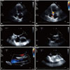
We report a case of a 56-year-old female on hemodialysis who underwent endovascular stenting to treat right subclavian vein thrombosis and experienced stent migration to the right atrium. She presented to the emergency department with complaints of chest pain. A 2-D transthoracic echocardiogram (TTE) was done as a part of the work up and demonstrated an unusual hollow mass in the right atrium with metallic echogenicity, in addition to a possible patent foramen ovale (Fig. 1, Supplementary movie 1). A transesophageal echocardiogram (TEE) was done for better evaluation of the right atrial mass and to rule out thrombus or vegetation (Supplementary movie 2). The TEE confirmed the presence of the migrated subclavian stent in the right atrium originating from the superior vena cava, with normal flow through it via color Doppler (Supplementary movie 3). It also showed a small patent foramen ovale seen in the inter-atrial septum, which was also confirmed with bubble study. The patient had a negative nuclear stress test, and her pain was attributed to musculoskeletal chest pain and responded to treatment with NSAIDs. As the patient was asymptomatic and the migrated stent did not have any subsequent clinical sequelae, the decision was made to proceed with the conservative approach and the stent was left in place. At her 3 months follow-up, she was still asymptomatic and a repeat TTE showed the migrated stent still in right atrium with no complications.
The cause of stent migration is not quite clear but poor insertion technique, excess mobility, turbulence, in addition to fibrosis and stenosis of the lumen by central venous catheters have been implicated.5)
If the migrated stent causes no immediate life threatening complications, the condition can be managed conservatively. However, valvular regurgitation, intracardiac thrombosis, embolization into the pulmonary artery branches and infection are potential complications and considered indications for intervention.6)
The migrated stents can be managed either percutaneously or by open surgery.7) Loop snares are the most widely used retrieval devices.8) Success rates of percutaneous techniques in the management of migrated stents exceed 90%.9) However, if percutaneous retrieval was not successful then open surgical methods should be used but they are associated with higher complications.

Figures and Tables

Fig. 1

A: Transthoracic echocardiogram apical four chambers view showing the displaced subclavian stent in the right atrium. B: Normal flow and no signs of turbulence within the displaced stent by color Doppler. C, D, and E: Transesophageal echocardiogram views with zoomed in imaging of the right atrium. At zero degrees the subclavian stent can be seen in short axis clearly (C). At 103 degrees angle the stent can be seen in long axis originating at the superior vena cava-right atrial junction with normal flow and no signs of turbulence by color Doppler (D and E). F: Transesophageal echocardiography showing contrast medium passing through the patent foramen ovale.

jcu-24-251-g001

References

1. Bagul NB, Moth P, Menon NJ, Myint F, Hamilton G. Migration of superior vena cava stent. J Cardiothorac Surg. 2008; 3:12.
2. Verstandig AG, Bloom AI, Sasson T, Haviv YS, Rubinger D. Shortening and migration of Wallstents after stenting of central venous stenoses in hemodialysis patients. Cardiovasc Intervent Radiol. 2003; 26:58–64.
3. Poludasu SS, Vladutiu P, Lazar J. Migration of an endovascular stent from superior vena cava to the right ventricular outflow tract in a patient with superior vena cava syndrome. Angiology. 2008; 59:114–116.
4. Dubois P, Mandieau A, Dolatabadi D, Chaumont P, Gurnet P, Thiriaux J, Delcour C, Creplet J. [Right ventricular migration of a stent after endovascular treatment of a superior vena cava syndrome]. Arch Mal Coeur Vaiss. 2001; 94:1180–1183.
5. Maintz D, Landwehr P, Gawenda M, Lackner K. Failure of Wallstents in the subclavian vein due to stent damage. Clin Imaging. 2001; 25:133–137.
6. Taylor JD, Lehmann ED, Belli AM, Nicholson AA, Kessel D, Robertson IR, Pollock JG, Morgan RA. Strategies for the management of SVC stent migration into the right atrium. Cardiovasc Intervent Radiol. 2007; 30:1003–1009.
7. Gabelmann A, Krämer SC, Tomczak R, Görich J. Percutaneous techniques for managing maldeployed or migrated stents. J Endovasc Ther. 2001; 8:291–302.
8. El Feghaly M, Soula P, Rousseau H, Chaiban F, Otal P, Joffre F, Cerene A. Endovascular retrieval of two migrated venous stents by means of balloon catheters. J Vasc Surg. 1998; 28:541–546.
9. Gabelmann A, Kramer S, Gorich J. Percutaneous retrieval of lost or misplaced intravascular objects. AJR Am J Roentgenol. 2001; 176:1509–1513.

Supplementary movie legends

Movie 1

Transthoracic echocardiogram apical four chambers view showing the displaced subclavian stent in the right atrium.

Movie 2

Transesophageal echocardiogram short axis view at zero degrees showing the subclavian stent in the right atrium.

Movie 3

Transesophageal echocardiogram long axis view at 103 degrees angle, the stent can be seen originating at the superior vena cava-right atrial junction with normal flow and no signs of turbulence by color Doppler.
TOOLS
Similar articles