

 PDF
PDF ePub
ePub Citation
 Print
Print


Introduction
Common atrium is a rare congenital heart defect comprising < 0.5–1% of all congenital heart diseases.1)2) Common atrium is characterized by complete absence of the interatrial septum, and is commonly accompanied by malformation of the atrioventricular valve.3) Most patients with common atrium experience symptoms during childhood. However, we describe a patient with common atrium who experienced his first obvious symptom at 48 years of age.
Case
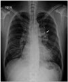
A 48-year-old man presented to the outpatient clinic with a 1-week history of intermittent palpitations. He had no significant medical history. On physical examination, wide splitting of S2 during auscultation was noted, but no other cardiac murmur was found. He had mild digital clubbing, but labial cyanosis at rest was not observed. Electrocardiography showed atrial fibrillation with rapid ventricular response. Laboratory data revealed an elevated hemoglobin level of 17.9 g/dL. Oxygen saturation obtained from pulse oximetry was 88%. Chest radiography showed mild cardiomegaly with a prominent left hilar shadow and increased pulmonary vascularity (Fig. 1).
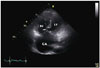
In order to evaluate the patient's atrial fibrillation, transthoracic echocardiography was conducted, which showed complete absence of the interatrial septum. At this point, we looked for an atrioventricular valve malformation, which frequently accompanies common atrium. The anterior common leaflet appeared to be attached to the crest of the ventricular septum by a chorda (Fig. 2), and a definite mitral and tricuspid valve cleft was not noticed. The right ventricle was markedly enlarged with mild pulmonary hypertension (estimated right ventricular pressure, 39 mm Hg).
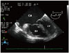
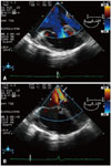
Because transthoracic echocardiography was slightly suboptimal, transesophageal echocardiography was conducted, which also showed complete absence of the interatrial septum. The anterior common leaflet appeared to be attached to the crest of the ventricular septum by a chorda (Fig. 3), as seen on transthoracic echocardiography. However, no interventricular shunt was found. In addition to right ventricular dilation with borderline hypertrophy, mild mitral and tricuspid regurgitation was observed (Fig. 4). These findings corresponded to common atrium with atrioventricular valve malformation, which is an atrioventricular septal defect with separate atrioventricular valves and an intra-atrial shunt only.
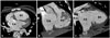
In order to check for anomalies of the systemic and pulmonary veins as well as accompanied cardiac anomaly, cardiac computed tomography was performed. The pulmonary veins drained to the left side of the common atrium, while the systemic veins drained to the right side. Visceroatrial, atrioventricular, and ventriculoarterial concordance were noted. There were no other accompanied anomalies (Fig. 5).
In order to evaluate the patient for genetic diseases, such as Ellis-van Creveld syndrome, which is related to common atrium, additional medical history taking and physical examination were conducted. On physical examination, characteristics of Ellis-van Creveld syndrome, such as polydactyly, short stature, hypodontia, and disproportionate distal limb shortening, were not observed. On additional medical history taking, he recalled experiencing a bit more shortness of breath than others during excessive exercise. He also had recurrent respiratory infections in childhood, but he did not see a physician because his symptoms were very mild. The patient is currently being treated with amiodarone and warfarin for atrial fibrillation, and is considering surgery to reconstruct the interatrial septum.
Discussion
Common atrium was first reported by Young and Robinson4) in 1907. Since then, common atrium occasionally has been reported as a cardiac component in patients with Ellis-van Creveld syndrome, trisomy 21, or heterotaxy syndrome with asplenia, whereas it is rare in nonsyndromic patients.3)5)6) The hemodynamic features of shunting in common atrium are very similar to those of a large atrial septal defect. However, in patients with common atrium, pulmonary and systemic venous blood tend to mix more easily, and systemic arterial oxygen saturation tends to be lower than in those with a large atrial septal defect.7) Consequently, most patients with common atrium experience such symptoms as exercise intolerance, exertional dyspnea, palpitations, frequent upper respiratory tract infections, cyanosis, and physical underdevelopment during childhood. Common atrium is frequently accompanied by anomalies of systemic venous drainage and atrioventricular regurgitation, which may increase mixing of blood and result in decreased systemic arterial oxygen saturation.2)7) Considering that increased mixing of arterial and venous blood can generate more severe symptoms at an earlier time, our patient is thought to have experienced relatively mild symptoms because he had no mitral and tricuspid valve cleft nor anomalies of systemic venous drainage, which are expected to increase regurgitation.
Because most patients with congenital heart disease show symptoms and receive a diagnosis before reaching adulthood, it can be difficult for physicians to consider the possibility of congenital heart disease in patients in whom symptoms are absent or well tolerated until late adulthood, as in the present case. Therefore, clinical suspicion is vital. If congenital heart disease, such as common atrium, is suspected, diagnosis can be made easily by using echocardiography. Our patient had been experiencing dyspnea during excessive exercise, and had such objective findings as digital clubbing and polycythemia, which increase the possibility of chronic hypoxemia. He also showed a prominent left hilar shadow and increased pulmonary vascularity, which can occur in patients with a left-to-right shunt,2)3) as well as cardiomegaly. Although he did not possess any anomaly related to Ellis-van Creveld syndrome, trisomy 21, or heterotaxy syndrome with asplenia, investigating the clinical characteristics of such diseases also may be useful for clinical suspicion.
The natural course of common atrium is poor. Later identification of common atrium increases the possibility of progression to arrhythmia, pulmonary vascular disease, or cardiac dysfunction. 5)6) Moreover, surgical correction can considerably slow progression of the disease.3) Therefore, early diagnosis is important even in well-tolerated cases.
Asuman Kaftan et al.8) reported a similar case of undiscovered common atrium in a 23-year-old patient. In addition, Demirelli et al.9) reported a case of incidentally diagnosed asymptomatic common atrium in a 23-year-old woman, while Hasanin and Kinsara10) reported a case of late-discovered common atrium in a patient with persistent left superior vena cava. However, to our knowledge, our case is the oldest patient with common atrium to have remained asymptomatic. Moreover, the present case shows that clinical characteristics found on physical examination, imaging studies, and laboratory tests can be useful for clinical suspicion of common atrium as long as physicians consider that patients with congenital heart disease can remain asymptomatic until late adulthood.
 XML Download