

 PDF
PDF ePub
ePub Citation
 Print
Print


Introduction
The speckle tracking echocardiography has been introduced as an advent to measure myocardial deformation with angle independent quantification of left ventricular (LV) twist. This technique is based on frame-by-frame tracking of natural acoustic markers that are generated by B-mode images. However there are very few data in pediatrics.
LV rotation, rotation rate, and radial displacement were directly measured using speckle tracking echocardiographic software from short axis views of the LV apex and base. Rotation was defined as circumferential rotation around the long axis of the left ventricle during systole, and rotation rate was the speed at which rotation occurred.
Traditionally, research in clinical cardiac mechanics involved analysis of short axis and long axis LV function and ejection fraction, but it has been advanced to three-dimensional ventricular deformation studies, including LV torsion.1)2) LV torsional deformation, based on helical myocardial fiber architecture, plays an important role with respect to LV ejection and filling performance.3)4)5)6) During the cardiac cycle, there is a systolic twisting and an early diastolic untwisting of the LV about its long axis due to opposite apical and basal rotations. The magnitude and characteristics of this torsional deformation are well established, and it is known that that LV rotation is sensitive to changes in regional and global LV function.7)8)9)10)11)12)13)14)15)16)17)18)19)
Therefore, interpretation of LV rotation represents a logical approach to quantifying LV function. However, there is no comprehensive study describing its normal development during childhood with respect to age-related change.
In systole, the LV apex rotates counterclockwise while the base rotates clockwise, creating torsional deformation originating in the dynamic interaction of oppositely wound epicardial and endocardial myocardial fiber helices.2) One of the special characteristics of static B-scan ultrasound imaging is the appearance of speckle patterns within the tissue, which result from constructive and destructive interference of ultrasound back-scattered from structures smaller than the ultrasound wavelength. Motion analysis by speckle tracking has been attempted using block-matching and autocorrelation search algorithms. Speckle motion has also been closely linked to underlying tissue motion when small displacements are involved.20)21)22)
A recently developed and noninvasive echocardiographic speckle tracking imaging (STI) technique, which is a novel ultrasound method for quantification of true two-dimensional (2D) heart motion independent of borders, Doppler or its beam angles, has been used for assessment and quantification of LV rotation and torsion.23)
LV torsion and untwisting show age-related increases in general, and when normalized by LV length, they demonstrate larger values in infancy and middle age. Notomi et al.24) has suggested that net LV torsion increases gradually from infancy to adulthood, but the determinants of this remain unclear.
The neonatal myocardium develops less force than that of the adult, and cardiocytes have been shown to increase both myofibrillar and sarcoplasmic reticulum contents after birth.24)25)26) Large changes in hemodynamic load occur during cardiac development and are associated with increased contractility resulting from alterations in the relative expression of sarcoplasmic protein isoforms.27) The giant sarcoplasmic protein 'spring' both resists passive stretch and helps the myocyte to recoil after contraction.28) In addition to these cardiac changes, arterial distensibility decreases from childhood to adulthood, which represents a stiffening of the arterial tree that increases afterload even in normotensive individuals.29)30)
In this study, we sought to investigate the alterations in LV torsional behavior from preschool age to school age in normal children.
Methods
Study participants
The participant children were recruited in the outpatient clinic from January to July 2014, when schedules to undergo clinically indicated echocardiography and from healthy volunteers. They were 2 to 14 years, without heart disease. The children with unstable hemodynamics and cardiac arrhythmias were excluded. They were divided into two groups: one group of twenty preschool-age children (2 to 6 years, mean age 4.5 ± 1.2) and the other group of twenty school-age children (7 to 14 years, mean age 10.5 ± 2.7).
Children were recruited from a group of pediatric patients referred for electrocardiography (ECG) or echocardiography to evaluate cardiac murmur, chest pain, and syncope. All subjects were normotensive and clinically well from a cardiovascular standpoint. Additionally, they showed normal sinus rhythm with a normal surface ECG and no structural or functional abnormalities on transthoracic echocardiography. They were free of past or present systemic disease.
Echocardiography
The main echocardiographic examinations were performed by one expert with Vivid 7 scanner (GE Vingmed Ultrasound, Horten, Norway) equipped with a phased-array transducer. Transducer frequencies, sampling rates, and sector width were adjusted for optimal speckle quality of the recordings, and LV short axis recordings were acquired. A 2D ultrasound STI analyzing software packages (Echopac PC, version 6.0, GE Healthcare, Horten, Norway) provided by the manufacture was used for offline analysis.
In this study, the proper short axis levels were defined at the basal level by the presence of the mitral valve and at the apical level by the LV cavity alone with no papillary muscles. The LV cross section was made as circular as possible. In each child, 3 consecutive cardiac cycles were acquired, and the data at end-expiration on the respiratory trace were selected.
The analyses were performed on a computer with customized software within the EchoPac platform (GE Medical Systems, Milwaukee, WI, USA).
Conventional echocardiograms were evaluated for LV systolic and diastolic function. After completion of standard comprehensive examinations to assess LV longitudinal myocardial motion, tissue Doppler imaging (TDI) analysis was performed offline, and the myocardial tissue velocity profile was obtained from an optimal measuring position set at the basal segment of the septum and LV lateral wall from apical four chamber projections. The mean frame rate was 150–180 frames per second, and the velocity range was 12–20 cm/sec to avoid aliasing for TDI acquisition. The measurements of maximal systolic and early diastolic velocities were obtained.
In addition, at the basal and apical short axis levels, radial transverse and circumferential strain values were obtained using the EchoPac program (GE Medical Systems).
LV rotation and torsion
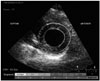
Spectral tracking echocardiography was performed for offline analysis, and LV rotation was then defined as angular displacement of the left ventricle about its central axis in the short-axis image (Fig. 1). These data were measured in degrees.
LV torsion was defined as the net difference of global LV rotation between apical and basal short axis planes at each time point and was calculated by the following equation:31)32)
Global torsion = apical global rotation - basal global rotation
Peak global torsion was defined as the maximal value of global torsion during the cardiac cycle.
Results
We divided the study population into 2 groups: twenty preschool-age children (2–6 years) and the other twenty school-age children (7–14 years). From conventional echocardiographic measures, LV ejection fraction (67.0 ± 2.0% vs. 66.5 ± 5.4%, p = NS) was not different between two groups.
LV rotation pattern
Apical rotation, which is consistently counterclockwise and is presented as a positive value, changed slightly from preschool age to school age without statistical significance, whereas basal rotation, which occurs in a clockwise direction and is represented by a negative value, changed significantly with aging (p < 0.05), especially at the inferior and septal segments (p < 0.02).
Global mean value of basal rotation was greater in preschool-age than in school-age children (-6.3 ± 3.0° vs. -4.4 ± 2.3°, p < 0.05). All of the six observed segments on short axis images, antero-septal, anterior, lateral, and posterior segments demonstrated a tendency of higher rotation in preschool-age children (antero-septal: -3.6 ± 2.5° vs. -2.6 ± 2.2°, anterior: -4.4 ± 2.4° vs. -3.4 ± 3.4°, lateral: -6.5 ± 2.8° vs. -5.6 ± 3.3°, posterior: -7.7 ± 4.0° vs. -5.9 ± 3.6°, p = NS, respectively), and inferior and septal segments exhibited statistically significant higher rotation in preschool-age children (inferior: -9.2 ± 3.5° vs. -6.6 ± 3.0°, septal: -8.0 ± 3.1° vs. -5.3 ± 3.6°, p < 0.02, respectively) (Table 1, Fig. 2).
Although there was no statistical significance, global mean apical rotation was also higher in preschool-age children (7.7 ± 5.1° vs. 6.8 ± 7.0°, p = NS). For the same six segments on short axis images, apical rotation data showed tendency of larger measurement in preschool-age children than in school-age children (antero-septal: 9.5 ± 4.5° vs. 8.0 ± 6.2°, anterior: 9.6 ± 5.1° vs. 8.1 ± 6.6°, lateral: 9.1 ± 5.6° vs. 7.7 ± 7.6°, posterior: 8.2 ± 5.3° vs. 6.5 ± 7.9°, inferior: 6.6 ± 5.5° vs. 6.1 ± 7.3°, septal: 8.4 ± 4.0° vs. 7.2 ± 7.0°, p = NS, respectively) (Table 1, Fig. 2).
Radial strain and circumferential strain with speckle tracking echocardiography
Basal radial strain was not different between segments (antero-septal: 31.5 ± 11.7% vs. 36.5 ± 18.6%, anterior: 40.3 ± 17.2% vs. 44.1 ± 19.8%, lateral: 54.1 ± 15.2% vs. 50.2 ± 20.5%, posterior: 58.7 ± 18.0% vs. 52.4 ± 23.7%, inferior: 53.3 ± 20.5% vs. 47.2 ± 24.4%, septal: 39.8 ± 18.3% vs. 32.2 ± 21.9%, p = NS, respectively). Apical radial strain showed statistically significant higher values in preschool-age children, especially at the anterior (52.8 ± 17.4% vs. 34.7 ± 23.2%, p < 0.02), lateral (55.8 ± 20.4% vs. 36.1 ± 22.7%, p < 0.02), and posterior segments (57.1 ± 17.6% vs. 38.5 ± 21.7%, p < 0.01) (Table 2).
Meanwhile, differences in basal circumferential strain were not statistically significant between segments (antero-septal: -26.7 ± 6.7% vs. -25.7 ± 8.2%, anterior: -13.9 ± 6.8% vs. -16.6 ± 5.7%, lateral: -17.4 ± 8.2% vs. -14.7 ± 5.7%, posterior: -18.3 ± 9.0% vs. -17.0 ± 6.3%, inferior: -24.6 ± 5.4% vs. -21.5 ± 7.2%, septal: -29.1 ± 6.2% vs. -25.9 ± 8.0%, p = NS). Apical circumferential strain did not show the significant difference between segments (antero-septal: -24.9 ± 4.7% vs. -25.5 ± 7.7%, anterior: -20.4 ± 6.8% vs. -20.8 ± 9.6%, lateral: -17.9 ± 7.2% vs. -18.4 ± 6.6%, posterior: -16.7 ± 6.4% vs. -18.4 ± 7.0%, inferior: -19.8 ± 4.3% vs. -21.0 ± 7.6%, septal: -23.1 ± 9.0% vs. -25.3 ± 7.8%, p = NS) (Table 3).
LV torsion pattern
With the torsion calculation from these basal and apical rotation data, LV torsion did not show the statistical difference between preschool-age children and school-age children. However, the preschool-age children had the larger measurements (12.6 ± 5.8°/cm vs. 9.5 ± 6.9°/cm, p = NS) (Table 4).
Discussion
Modulation of LV torsion appears to reflect myocardial mechanical maturation in childhood, which is influenced by contractility, loading conditions, and possible myogenetic changes through children's growth in life.
In this study, all forty enrolled children (2- to 14-year-old) showed no significant difference in LV ejection fraction. Basal rotations in inferior and septal wall significantly increased in preschool-age children. Otherwise, without statistical significance, basal and apical rotations of preschool-age children were higher than those of school-age children. From these rotation data, calculated LV torsion was larger in preschool-age children, without statistical difference. These results can suggest that myocardial activity might be more dynamic with the larger rotation and torsion in younger preschool-age children in comparison to older school-age children. Notomi et al.24) suggested that LV torsion was higher in infants (n = 9, 9 ± 11 month, < 2 year) than in older children (n = 8, 7 ± 3 year), adolescents (n = 8, 16 ± 2 year), and young adults (n = 10, 28 ± 3 year), which correlates with the finding that contractility is higher in children under 2 years of age due to higher metabolic demand in comparison to older children.33) Although we did not include infants in this study, it is possible that younger preschool-age children demonstrated higher torsion for the same reasons that infants exhibit higher contractility. Meanwhile, the right ventricles of newborn infants are hypertrophied compared with those of older school-age children and adults due to the systemic pressure and resistance of the right ventricle. Infants may have relative LV hypertrophy as well, as previously presented by Harada et al.34) This hypertrophy recedes with a concomitant change in myofibril architecture with growth and aging, which may yield the lesser ventricular rotation.
This is the foremost study to measure rotation and short axis radial and circumferential strain together at the base and apex in order to observe how these parameters interact in children.
Strain measure for myocardial deformation in radial and circumferential directions showed no statistical difference at the base with increase of age, which correlates well with the report that LV geometry and systolic ejection fraction are constant from infancy to adulthood.33)35) However, the basal rotation was greater at the inferior and septal segments in younger preschool-age children (Table 1), which was caused from noticeable tendency of higher strain at the exact inferior and septal segments in radial and circumferential directions (Table 2 and 3). Perhaps at base level, both of the radial and circumferential myocardial fibril may affect LV rotation and torsion with equivalent importance.
Meanwhile, the apical radial strain at anterior, lateral, and posterior segments were significantly higher in preschool-age children (Table 2); however, apical circumferential strain was not different between the two groups at anteroseptal, anterior, lateral, posterior, inferior, or septal segments on short axis images (Table 3). Even though the higher apical rotation values seen in preschool-age children were not statistically significant, these values might be affected much more by circumferential deformation rather than radial deformation (Table 1). The fact that apical circumferential strain was found to be greater than radial deformation may have an impact on apical rotation and torsion. At the apex, circumferential myocardial deformation may be more important for myocardial performance.
We observed that rotation did not change much between 2 to 14 years old, while the age-related decrease in LV torsion during childhood resulted from a subtle change in radial and circumferential strain of basal and apical segment myocardial deformation.
In terms of future clinical impacts, having normal control reference values for children's LV torsion could be useful for assessing the status of various myocardial diseases. LV torsion might be a useful measurement of cardiac performance, which may allow for better understanding of the myocardium in the following conditions: cardiomyopathy, hypertension, postoperative congenital heart disease, and other myocardial changes. Furthermore, this systolic torsion study may provide new insight into the mechanistic manifestation of diastolic characteristics in childhood growth.
The limitation of this study is very small sample size, and including some of subclinical patient group for normal children with reasonable ejection fraction, such as chest pain group and syncope.
In conclusion, rotation values were found to be higher tendency in preschool-age children than in school-age children. We observed a decreasing trend in rotation and torsion values with increasing age from 2 to 14 years old. Although there was no statistically significant age-related change in LV rotation between these two groups, the decreasing trend in rotation and torsion twist values during childhood warrants further investigation.


 XML Download