

 PDF
PDF ePub
ePub Citation
 Print
Print


A 71-year-old female patient was referred for shock cardioversion following diagnosis of lone atrial fibrillation.
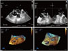
Transesopageal echocardiography was performed as routine workup. Interestingly a circular membrane like structure in the left atrial appendage (LAA) was observed (Fig. 1A, Supplementary movie 1). Using 2D X plane imaging echocardiography, where an orthogonal view can be acquired through the midline of a primary image and displayed as a secondary image, an extra thin LAA wall was evidenced showing sigmoid anatomy (Fig. 1B). Local pericardial effusion was questioned implicating that this specific structure accounted for the thin LAA wall. 3D echocardiography evidenced the entrance of LAA, surrounded by the pericardial wall at a distance, due to the presence of pericardial effusion (Fig. 1C and D, Supplementary movies 2 and 3). The patient underwent uneventful direct current shock cardioversion and remains in sinus rhythm at nine months follow up.
Cardiac magnetic resonance imaging confirmed the diagnosis of local pericardial effusion in the LAA area. No structure, thrombi or membrane were documented into the LAA (Supplementary movie 4).
Local pericardial effusion in the LAA area is an extremely rare finding.1)2) It is clinically important to be aware of this benign clinical finding that should be differentiated from LAA obstructive or non-obstructive membranes or thrombi, especially in patients with atrial fibrillation undergoing cardioversion or planned for specific transcatheter therapies such as implantation of LAA closure devices.3)4)5)
 XML Download