

 PDF
PDF ePub
ePub Citation
 Print
Print


Introduction
Lipomatous hypertrophy of the interventricular septum is extremely rare and it usually affects interatrial septum.1) There are limited reports of this entity in the literature2)3) however in our knowledge is the second case diagnosed by magnetic resonance imaging (MRI).4) Lipomatous hypertrophy should be differentiated by lipomas in the context that the latter are usually encapsulated and round-shaped while they do not exhibit myocardial fiber infiltration.4)
Although lipomatous hypertrophy has been associated in the past with sudden death,5) there is no enough evidence to support any kind of medical treatment or action in the case of lipomatous hypertrophy of the interventricular septum. This condition has been associated in some reports with atrial or ventricular arrhythmias following variable degree of conduction system infiltration. Additionally in cases of severe interventricular septal hypertrophy there might be left ventricular outflow obstruction and in this case surgical rejection might be a reasonable management.
Case
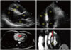
A 46-year-old asymptomatic male with no medical history, was referred for routine cardiology examination due to his frequent participation in competitive sports. On physical examination his blood pressure was normal, his heart rhythm was regular and no heart murmurs were detected. The patient's electrocardiogram showed a mild sinus tachycardia of around 100 beats per minute, an incomplete right bundle branch block pattern and no evidence of left ventricular hypertrophy (Supplementary Fig. 1). His echocardiographic study revealed a hyperechogenic interventricular septum, with a measured thickness of 1.34 cm, suggestive of asymmetrical left ventricular hypertrophy (Fig. 1). There was no left ventricular outflow obstruction in rest neither following moderate treadmill exercise and there were no evidence of mitral valve regurgitation. His 24 h Holter monitoring documented rare monomorphic ventricular extrasystoles and the patient was further referred for cardiac MRI, in order to evaluate possible myocardial fibrosis indicative of hypertrophic cardiomyopathy.
MRI showed extensive fatty infiltration of the interventricular septum. This area of lipomatous hypetrophy was nonhomogenic, not well circumscribed and was clearly defined from the rest normal myocardium (Fig. 1). Furthermore there was evidence of some infiltration by myocardial fibers. Right ventricular myocardial wall had a normal structure and appearance.
In the case we are presenting, there was no overt symptomatology, the 24 h Holter monitoring showed only rare ventricular extrasystoles and the exercise treadmill test was uneventful. Additionally since there was no echocardiographic evidence of left ventricular outflow obstruction no further management was discussed. Avoidance of competitive sports and yearly routine follow-up was finally recommended for this specific patient.
Discussion
Asymmetrical septal ventricular hypertrophy might be secondary to septal lipomatous hypertrophy or encapsulated lipomas. Typical echocardiographic features of ventricular septal lipomatous hypertrophy is the characteristic septal hyperechogenicity in the long axis and four chamber view which is not associated with mitral valve leak, systolic anterior motion of the valve or left ventricular outflow obstruction. Cardiac MRI is the preferred imaging modality for diagnosis of this specific situation, as it shares significant sensitivity for myocardial fat infiltration identification. In some rare cases endomyocardial biopsy is needed in order to establish the diagnosis.
 XML Download