

 PDF
PDF ePub
ePub Citation
 Print
Print


Introduction
Unileaflet mitral valve is the rarest form of congenital anomalies of the mitral valve. True unileaflet mitral valves are usually associated with severe mitral regurgitation (MR). This valvular defect is often detected in infancy and tends to be incompatible with life. In asymptomatic patients, the presence of a unileaflet mitral valve is most commonly due to a severely hypoplastic posterior mitral leaflet.
Case
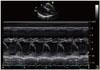
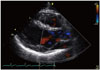
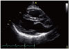
A 22-year-old otherwise healthy man presented to the clinic complaining of recurrent episodes of "heart racing". Physical examination was unremarkable. Prior laboratory testing was not significant for anemia, infection, thyroid or adrenal disease, vitamin deficiencies or electrolyte derangements. He underwent serial electrocardiograms and holter monitoring and had no detection of cardiac arrhythmias. A transthoracic echocardiogram revealed a preserved left ventricular ejection fraction of 55%, normal left ventricle size and thickness, normal diastolic filling and an unusual incidental finding of a unileaflet mitral valve. The anterior mitral valve leaflet was markedly elongated, mildly thickened, and occupied the entire closure line of the mitral annulus with no contribution of the hypoplastic posterior leaflet. The coaption zone was intact with no MR (Fig. 1, 2, and 3, Supplementary movie 1 and 2). As to his palpitations, he was diagnosed with an underlying anxiety disorder and successfully treated with anxiolytic therapy. Follow up was arranged yearly for his abnormal mitral valve.
Discussion
Congenital malformations of the mitral valve are extremely rare and include congenital mitral stenosis, parachute mitral valve, double orifice of the mitral valve, cleft mitral valve, atresia, and unileaflet mitral valve.1) Unileaflet mitral valve is the rarest of these congenital anomalies. Aplasia of the posterior mitral valve leaflet is responsible for unileaflet mitral valve in the neonatal period and associated with severe MR that is usually incompatible with life without surgical intervention.2) Existing literature in the field document cases of unileaflet mitral valve in asymptomatic patients of all age groups and mostly result from a severely hypoplastic posterior mitral valve.1)2)3)4)5)6)7) The prevalence of this anomaly is estimated to be 1:8800 in German population;2) however, the true prevalence is expected to be much lower in the general population.
To the best of our knowledge, there have only been 11 reported cases of post-neonatal unileaflet mitral valve in the literature, of which 70% of cases were found to be in asymptomatic patients. The majority of cases were found after detection of a systolic murmur during routine physical examination.1)2)3)4)5)6)7) In the aforementioned cases, transthoracic echocardiography (TTE) revealed a thickened and elongated anterior mitral valve with significant prolapse into the left atrium.2) In two of the cases, patients developed severe MR and underwent surgical correction and were found to have hypoplasia of the posterior mitral valve with absence of the chordae tendinae.4)6) It was also suggested that 3D echocardiography with transesophageal echocardiography (TEE) outlines the congenital abnormalities of mitral valve better than TTE or only TEE.1)
The prognosis in asymptomatic individuals is unclear; however, there is a potential for worsening MR with age due to annular dilation. There are no guidelines or recommendations currently for screening or follow-up monitoring of these patients. In our opinion, it may be appropriate to follow-up such patients with annual physical examinations and perform echocardiography if symptoms develop or MR murmur develops/worsens.
In conclusion, hereby we represent a rare case of unileaflet mitral valve and review not only the importance of echocardiography in accurate diagnosis, but also highlight the importance of periodic surveillance with echocardiography in early detection of MR in such cases.
 XML Download