

 PDF
PDF ePub
ePub Citation
 Print
Print


Introduction
The incidence of ischemic heart disease is lower in women than that of men but it is steadily increasing in Korea partly because of increased longevity in women.1) For proper management of women ischemic heart disease, early detection of women coronary artery disease (CAD) is crucial, but clinicians commonly confronts difficulty because of ambiguous clinical presentations of women CAD.2)3) Moreover the diagnostic accuracy of exercise-stress electrocardiography (ECG), which is recomemded as an initial diagnostic test for CAD, is reported to be lower than men.4)5) The exercise induced ST segment depression has been known to have low specificity as a marker of myocardial ischemia because of higher frequency of abnormal baseline ECG or low electrocardiogram voltage in women and digoxin like effect of estrogen.5)6) Another point that makes lower accuracy of exercise test is women commonly have difficulties in performing optimal exercise level. As the initiation of coronary disease in women is later in their life than men, many women patients have associated comorbidity or musculoskeletal problems to reach sufficient exercise level.7)
Because of above mentioned shortcomings of exercise-stress ECG, it has been suggested that exercise or pharmacologic stress echocardiography may be better noninvasive tests to diagnose CAD in women.6)8)9) But in Korea, the diagnostic utility of stress echocardiography has not been clearly investigated in large number of cases and reported publications were mostly focused to male gender. And the diagnostic accuracy of stress echocardiography was not evaluated by gender difference.10)11)12)13)
The aim this study was to evaluate the diagnostic accuracy of dobutamine stress echocardiography (DSE) for detecting fixed coronary artery stenosis (CAS) in comparison with exercise-stress ECG test in Korean women who presented with chest pain.
Methods
Study population
Study populations were consisted by 225 female patients who had taken DSE from the Korean women's chest pain registry (KoROSE). KoROSE study is a multi-center study led by Women Heart Disease Research Working Group in Korea aiming to investigate the clinical characteristics of women who presented with chest pain in outpatient cardiology clinic. Women were eligible for the study if they; 1) had a history of typical/atypical chest pain or ischemic equivalent symptoms; 2) were aged ≥ 20 and less than 80 years; 3) were capable of performing treadmill exercise test (TET); 4) had no significant structural or valvular heart disease; 5) had neither angiographically confirmed previous CAD nor prior myocardial infarction. Patients with end-stage renal disease, chronic obstructive lung disease, primary pulmonary hypertension, and autoimmune disease were excluded. This prospective sub-study was performed at 4 cardiology centers out of total KoROSE participating 20 centers from October 2011 to December 2014 in a consecutive manner. Exercise stress ECG test and DSE were performed in each patient within a month before coronary angiography (CAG). Based on guideline,14)15) pretest probability was evaluated in each patient by age and symptom and the patients were classified as low (< 20%) imtermediate (20–80%) and high probability (> 80%). The presence of hypertension, diabetes, and hyperlipidemia was defined if patients had been diagnosed before by their physician or had taken antihypertensive, antidiabetic, or antihyperlipidemic drugs. Each center obtained participant approval and Institutional Review Board approval for the study prior to enrollment.
Exercise stress ECG
TET was performed according to standard Bruce protocol. The symptoms and blood pressure (BP) were monitored throughout the exercise test. Twelve lead ECG at baseline at the end of each stage, at peak exercise, at 1 minute after exercise and every 2 minute at recovery period were recorded. Functional capacity of each patient was evaluated by metabolic equivalents (METs). Duke treadmill score (DTS) was calculated based on exercise duration, degree of ST segment shifting and presence of angina symptom during exercise.16) The patients were classified as previously reported system using DTS: low risk-DTS ≥ 5, moderate risk-DTS from 5 to -11, high risk-DTS ≤ -11.16)17)
TET was terminated by any of following reasons:14) 1) Heart rate (HR) achievement more than 90% of age predicted maximal HR without any symptoms (220 beats per minute–age). 2) Increased systolic BP (SBP) over than 250 mm Hg. 3) Intolerance to exercise due to typical chest discomfort, dyspnea or fatigue. 4) Ischemic ST changes, 5) significant arrhythmia. The ischemic ECG changes were defined if horizontal or down-sloping ST segment depression was developed more than 1 mm at 60 to 80 ms after the J point was developed more than 1 mm in leads without pathological Q waves in more than 2 leads.18) If baseline ECG was shown abnormalities affecting ST segment like bundle branch block, left ventricular hypertrophy, ischemic change was defined that the calculated difference from rest to exercise of changes was more than 1 mm.19) The definition of inadequate study was that maximal HR was not increased less than 85% of age predicted maximal HR without development of ischemic ST change.
Dobutamine stress echocardiography
At the day of stress echocardiography, beta blocker (BB), calcium channel blocker (CCB) and nitrates were withheld. The patient fasted for over 6 hour before the test. After baseline image acquisition, dobutamine was administered by the continuous intravenous infusion at the doses of 10, 20, 30, and 40 µg/kg/min for 3 min each, and then atropine was injected if target HR (85% of age predicted maximal HR) did not achieved. The atropine was administered as repeated 0.5 mg boluses up to total dose of 2 mg if HR increment was inadequate. The presence of any symptoms, BP, and ECG were monitored during examination. The infusion of dobutamine was discontinued if one of the following was occurred: age predicted target HR achieved, anginal symptoms, ventricular or supraventricular tachycardia, numerous extrasystoles, sinus bradycardia, atrioventricular block, hypotension (SBP < 90 mm Hg), a drop of SBP exceeding 30 mm Hg, or an increase of BP exceeding 220/120 mm Hg. Parasternal long axis, short axis at papillary muscle and 3 apical views were recorded at baseline, each stage of dobutamine dose and recovery periods. The wall motion of left ventricle was interpreted by 17 segments and wall motion score index was calculated, as previously described.20) The positive finding was defined as development of new wall motion abnormality or worsening wall motion abnormality. Peak velocity and pressure gradient on left ventricular outflow tract (LVOT) in each stage were evaluated by pulsed wave and continuous wave Doppler interrogations. Dynamic LVOT obstruction (DLVOTO) was defined as the existence of a dagger shape systolic flow with pressure gradient > 35 mm Hg in the LVOT or mid-ventricular region which was not present at baseline and disappeared at recovery.
Coronary angiography
CAG was conducted by standard methods. 2–3 images for right coronary artery and 3–5 images for left coronary artery were recorded. The severity of CAS was assessed visually by an intervention cardiologist. The significant CAS was defined as > 50% luminal narrowing of at least one coronary artery. Severe CAS was defined as ≥ 75% luminal narrowing on more than one coronary artery.
Statistical analysis
Quantitative data were expressed as mean ± SD and categorical data was presented as frequency and proportion. Independent t and chi-square tests were used for comparison between groups of continuous and categorical variables, respectively. The sensitivity, specificity, and positive predictive value (PPV) and negative predictive value (NPV) of TET and DSE for detecting CAD were calculated by the usual fashion. The diagnostic accuracy was calculated as the number of patients (true positive + true negative) / total patients. SPSS version 20.0 for Window (SPSS Inc., Chicago, IL, USA) was used for statistical analysis.
Receiver operating characteristic (ROC) curve analysis was used to obtain predictive power of positive finding on DSE, ischemic ST change of TET and DTS. The comparison of ROC curves was performed by using MedCalc for Windows version 9.2.0.1 (MedCalc Software, Mariakerke, Belgium).
Results
Baseline characteristics
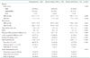
Among 225 patients who were recruited in comparison study, 23 patients were excluded for comparison analysis either because of inability to perform TET by orthopedic problem or incompletion of dobutamine infusion stress due to intolerable symptoms. Baseline characteristics of remaining 202 patients who underwent both tests are presented in Table 1.
Significant CAS was detected in 54 patients (26.7%). Among 54 patients, 27 patients had single-vessel CAS and 27 patients had multi-vessel CAS (2-vessel CAS: 9 and 3-vessel CAS: 18 patients). Severe CAS was present in 43 patients. The mean age of study population was 58.1 ± 10.2 years and patients with significant CAS was older than patients without CAS (64.7 ± 9.5 vs. 57.0 ± 9.7, p < 0.001).
The mean of pretest probability of total study population was 47.8 ± 30.6%. About 70% of study population belonged to higher than intermediate probability group and 30% were low pretest probability group. The portion of low pretest probability subjects was higher in patient without CAS than that of patients with CAS as expected (Table 1). In patients with CAS, the prevalence of hypertension and diabetes were higher than patients without CAS. Though prevalence of hypertension were higher in the patients with CAS than in patients without CAS, the percent of angiotension converting enzyme inhibitor, angiotension receptor blocker, BB or CCB taken by patients were similar between two groups.
Treadmill exercise test
TET data were presented in Table 2. Exercise time and exercise METs were shorter and lower in patients with CAS than patients without CAS. 34 patients prematurely terminated exercise before achievement of 85% age predicted maximal HR. 10 patients stopped exercise due to fatigue, 13 patients due to dyspnea, 9 patients due to leg patin and 2 patients due to combination of chest pain and ischemic ST change. The number of patients who achieved ≥ 85% of age adjusted maximal HR was higher in patients without CAS (n = 124, 83.8%) than in patients with CAS (n = 32, 59.3%). Baseline and exercise SBP higher in patients with CAS than without CAS.
Baseline normal ECG without ST segment abnormality was observed in 190 patients (94.1%) and left ventricular hypertrophy with ST segment change or right bundle branch block were present in 7 and 5 patients respectively. There was no difference in percent of normal or abnormal baseline ECG findings between patient with CAS or not (Table 2). No patient had left bundle branch block (LBBB) and ST segment elevation at baseline.
Ischemic ST change with typical pattern of ischemic ST depression developed more frequently in patients with CAS (53.7% vs. 26.4%, p < 0.001). Overall, the sensitivity and specificity of treadmill exercise ECG were 53.7% and 73.6%, respectively. When data was analyzed after excluded the patients who stopped the exercise prematurely, the test sensitivity was increased to 70.7% with 69.3% specificity.
The mean of DTS was also significantly lower in patients with CAS than that of patients without CAS (-0.66 ± 7.3 vs. 5.27 ± 5.1, p <0.001). As classified by DTS, 63% of patients with CAS was belonged to moderate risk group, while the majority of patients (62.8%) without CAS were classified low risk group.
Dobutamine stress echocardiography
In total study population, the administered average peak dobutamine dose was 37.7 ± 5.4 µg/kg/min without difference between paients with and without CAS (38.1 ± 4.7 µg/kg/min vs. 36.6 ± 6.8 µg/kg/min, p = 0.14). Atropine was used in 115 patients (57%). Test was prematurely terminated in 12 patients (5.3% of 225 patients) because of high BP (n = 3), ventricular tachycardia (n = 2) and intolerable symptoms such as nausea, headache and chest pain which was not accompanied by abnormal regional wall motion (n = 7).
Among 202 patients who undertaken both tests, 9 patients without CAS (6.1%) and 10 patients with CAS (18.5%) had minor regional wall motion abnormality at baseline (Table 3). And it was worsened in 2 patients without CAS and 8 patients with CAS, respectively. In 183 patients with normal regional wall motion at baseline, new regional wall motion abnormality developed in 36 patients. Therefore inducible ischemia was detected in 46 patients (22.8%) in patients who complete dobutamine stress test. The overall calculated test sensitivity was 70.4% and specificity was 94.6%. Test sensitivity of DSE was increased according to the number of stenotic epicardial coronary artery (51.9% for 1-vessel, 77.8% for 2-vessels, and 94.4% for 3-vessel CAS, p = 008). The sensitivity and specificity for localization of coronary stenosis by DSE was 55.9% and 91.3%, respectively.
The worsening of regional wall motion was observed in 8 patients of who had no significant coronary stenosis. These deteriorations of wall motion were localized to posterior (n = 4) and inferior wall (n = 3). Compared to true negative patient, these patients with false positive had higher SBP at peak stress than true negative patients (148.4 ± 21.7 vs. 130.9 ± 23.9, p = 0.046). They also had thicker ventricular wall and lower early diastolic mitral annulus velocity than patient with true negative (4.9 ± 1.7 vs. 6.7 ± 1.9, p = 0.015).
DLVOTO developed after dobutamine infusion in 64 patients (31.7%). The prevalence of DLVOTO or peak velocity were similar in both patient group.
Comparison of TET and DSE for detection of CAS
The diagnostic accuracy of TET and DSE are compared in Table 4. As described previously in each test result, DSE had superior sensitivity specificity, PPV and NPV than TET in diagnosing coronary stenosis by either ≥ 50% and ≥ 75% coronary stenosis criteria. When patients who did not perform adequate exercise level during exercise ECG without ischemia induction were excluded (non-diagnostic ECG due to inadequate stress level, total 34 cases), the sensitivity of TET became similar to DSE but specificity was still lower (Table 4). In patients with multi-vessel CAS, the sensitivity and specificity of both test increased but the trends that DSE had higher test accuracy than TET was remained (Table 5).
The clinical usefulness of stress test is highest in the patients with intermediate pretest probability and well documented in present guideline.19) Therefore test accuracy in patient with intermediate pretest probability was analyzed and presented in Table 6. In these group also, the sensitivity and specificity of DSE were higher than TET for CAS detection. And in patients who could not achieve ≥ 85% age predicted maximal HR on TET, DSE had a substantial specificity and diagnostic accuracy for predicting of CAS.
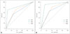
As the DTS has been known to have a greater predictability of CAS especially in women, ROC curve analysis was performed including TET, DTS, and DSE in detecting coronary stenosis (Fig. 1). DSE showed higher area under curve (AUC) [0.825, 95% confidence interval (CI) 0.765–0.875, p = 0.033] than TET (0.637, 95% CI 0.566–0.703, p = 0.039) and DTS (0.688, 95% CI 0.619–0.751, p = 0.04) for the prediction of CAS (Fig. 1A). And for the prediction of multi-vessel CAS, DSE had the higher test AUC (0.886, 95% CI 0.834–0.927, p = 0.014) over the TET (0.677, 95% CI 0.608–0.741, p = 0.05) and DTS (0.709, 95% CI 0.641–0.771, p = 0.032).
Discussion
This is the first comparison study of TET and DSE in Korean women who presented with chest pain in outpatient clinic. Present study demonstrated that DSE had a higher diagnostic value than TET in Korean female patients by the head to head comparison in each patient. As the disease severity of CAS worse, DSE had greater diagnostic accuracy than TET and these trends remained similar when the data was analyzed in patients with intermediate pretest probability before TET or DSE.
For the clinician, the diagnosis of CAD in women is challenging because of atypical clinical presentations and suboptimal test accuracy of exercise ECG test which is recommended as an initial test in women who suspected coronary disease.5) It has been known that the exercise test sensitivity and specificity is lower in women than men.4)21) Because of these reasons, adding imaging modality to detect myocardial ischemia after stress may be more practical way to diagnose CAD in women. 22)23) But there has been no data regarding accuracy of initial diagnostic test for CAD in Korean female patients. In has been known that the prevalence of obstructive CAD was lower in Asian than in Western, especially in women.24)25)26) Moreover compared to western women, Asian women presented more atypical symptoms in patients with stable angina.27) Because clinical characteristics and risk factors of CAD were different between Asian and Western female patients, the accuracy of noninvasive diagnostic tests and its clinical applications need to be evaluated in Korean female patients.
In present study, the test sensitivity and specificity, PPV and NPV were much higher with DSE than that of TET. When patients who did not achieve adequate HR during exercise were excluded, the test sensitivity of TET increased which was comparable to sensitivity of sensitivity. But still the specificity and test accuracy of TET were much lower than that of DSE. In various criteria of CAS and categorization of study subjects, the test accuracy was higher with DSE than TET. Therefore, the diagnostic utility of TET appeared limited and imaging stress test should be considered for the detection of CAD in Korean women who presented with chest pain in outpatient clinic. Especially the inadequate exercise level of stress seriously diminished test sensitivity, which was observed in 23% of present study population, therefore test with combination of non-exercise stressor and imaging modality may be an alternative to TET as an initial diagnosing method in women. For this purpose, DSE appeared to be a useful diagnostic method especially in deconditioned female patients. Although the PPV of TET was bad and the NPV of TET was relatively good, this information could be used in clinical decision process in physically active patient.
The sensitivity of both tests to diagnose CAS in this study appeared lower than previously reported values.4)9) The possible explanation for low sensitivity of both tests may be the low prevalence and less severity of significant CAS in study population. In present study, the prevalence of CAD was 26.7%. The prevalence of CAD is low compared with previously reported values.28) Lower prevalence of CAD in study population could lower the test sensitivity and specificity because of noninvasive nature of diagnostic method. Another point to be addressed is only 50% of patients with CAS had multi-vessel CAD. High proportion of single-vessel coronary disease in this study subjects may decrease the test sensitivity also. From these possibilities, when both test sensitivity and specificity were tested for detecting multi-vessel stenosis, the sensitivity of TET increased from 53.7% to 59.3%, and 70.4% to 88.9% in DSE. Thus the test result of noninvasive test need to be interpreted by referring the clinical characterization of tested patients also.
Besides of higher test accuracy, DSE has several advantages over TET. Localization of ischemia is possible during ischemia induction.29) Quantitation of ischemia extent and detection of ischemia induced valvular dysfunction can be detected during DSE also. In this study, the sensitivity to localize coronary artery territory by DSE was 55.9% with 91.3% specificity which was lower sensitivity than that of previous study.28)30)31) But the incidence of CAS in present study population was very low and test accuracy may change by few cases, the simple comparison may not be appropriate. Another advantage of DSE over TET is test feasibility. In this study, 10% of participating women patient could not exercise at all. Another 23% of patient who tried to do exercise test did not reach adequate exercise level to interpret the stress ECG test properly. But DSE was possible in 95% of total study patient safely.
The 9.4% of study population had abnormal regional wall motion at baseline. The possible explanation of baseline wall motion abnormality were hibernating myocardium due to critical coronary stenosis or misinterpretation of wall motion abnormality by visual analysis.
The reported false positive DSE test rate was about less than 15%.32) In present study the false positive rate was 5.4%, therefore the quality of DSE was comparatively acceptable. The well-known related factors of false positive DSE, such as LBBB, prior myocardial infarction and hypertensive response to exercise (typically SBP over 220 mm Hg),33) might have not relevance to false positive case in present study.
Additional useful finding during DSE is DLVOTO which was observed in 64 patients (31.7%) in this study. It was higher than previously reported value.34)35) Because of smaller LVOT size is prone to develop DLVOTO during increased inotopic status, smaller chamber size of women may be one reason for high incidence of DLVOTO in this study.36) It had been reported that, development of DLVOTO is related with the exercise intolerance and ischemic symptoms37)38) in patient without CAS, this finding may useful to understand the patient's symptoms. But its clinical and prognostic implications need to be investigated further.
Present study had several limitations. First angiographic evaluation was assessed only diameter stenosis of coronary artery. Functional assessment of coronary stenosis was not assessed. And myocardial ischemia caused by microvascular dysfunction which is not common but unique in female patient has not been assessed. Another limitation was inherent disadvantage of DSE. The performance of DSE was not possible in every participating hospital for KoROSE study. DSE is operator dependent test and needs certain degree of experience. And diagnostic power of DSE was decreased in patients with suboptimal image quality. This should be partially overcome by including deformation image analysis or by administration of contrast during DSE.39)40) All DSE was interpreted at core lab and no study was excluded because of suboptimal image. It could be possible that the researcher in each center may select the patient with optimal baseline image quality and may cause selection bias. However prevalence suboptimal image is much less in Asian patients than Western patients because of low incidence of obesity, effect of selection bias may be minimal.
In conclusion, the present study showed higher sensitivity and specificity of DSE than TET for the detection of CAS in Korean female patients who presented with chest pain with good feasibility. The diagnostic accuracy of TET was suboptimal but the negative ECG findings after exercise stress could be useful in excluding CAS. From these findings, we conclude that DSE could be used as the first-line test to diagnose CAS in selected patients who presented with chest pain.


 XML Download