

 PDF
PDF ePub
ePub Citation
 Print
Print


Introduction
Left ventricule (LV) systolic contractile dysfunction plays key roles in the pathophysiology of decompensation after acute myocardial infarction (AMI) or heart failure causing irreversible cardiac remodeling.1)2)3)4)5) It has been widely studied that cardiac remodeling predicts adverse clinical outcome.6)7)8) We have many studies focusing on left ventricular ejection fraction (LVEF) for its prognostic value for clinical outcome in patients 134with heart failure or myocardial infarction.1)2)3)4)5)8)9)10)11) The process from ST elevation myocardial infarction (STEMI) to LV remodeling is well documented in previous studies.6)7) Recent breakthrough of 2D speckle tracking analysis provided a new non-invasive methodology to assess cardiac functioning.12)13) 2D speckle analysis is mainly automatic but it includes some manual works which needs specialist to minimize intra- and inter-observer variability. Through extensive research, the validity of multidirectional strain analysis has been confirmed.14)15) The featuring parameters of 2D speckle tracking analysis include global longitudinal strain (GLS), global circumferential strain (GCS) and torsion. Those parameters independently reflect complex cardiac 3D contraction. Many studies have agreed on the fact that GLS and GCS correspond to subendocardial and mid-wall functions, respectively. These parameters are associated with cardiac viability,16)17) and adverse outcome.18)19)
In this study, we sought to investigate which echocardiographic parameters will be the best predictors of LV remodeling after STEMI.
Methods
Patient characteristics
Between July 2009 and May 2012, 354 STEMI patients were recruited from Seoul National University Bundang Hospital in South Korea. High risk patients, those who were older than 85 years old, who had atrial fibrillation, who underwent emergent coronary artery bypass graft (CABG) after coronary angiography (CAG), who require mechanical cardiac support such as intra-aortic balloon pump (IABP) or extra-corporeal membrane oxygenation (ECMO), who are co-morbid with cardiogenic shock, who died in hospital were excluded from the study (total 45 patients). Of these, 239 patients had follow up echocardiography at least 6 months interval. We further excluded patients with poor image qualities and therefore total 208 STEMI patients were evaluated in this study. The diagnosis of STEMI was made by electrocardiography findings with typical symptoms, which was to be confirmed by elevated cardiac enzymes such as CK-MB and troponin I.
All patients were successfully reperfused by either percutaneous coronary intervention (PCI) or thrombolysis. Then, 2D echocardiography was performed within 24 hours of those treatments. Before the immediate revascularization at admission, the time taken from door to balloon time was measured. During the PCI, infarct lesion(s) and culprit artery were assessed through CAG. The following baseline clinical and demographic data were obtained retrospectively from hospital electronic medical record (EMR): age, sex, weight (kg), height (cm), body surface area (m2), presence of hypertension, presence of diabetes, current smoking status, previous history of PCI or CABG, systolic blood pressure, and diastolic blood pressure.
Lab data were also obtained from EMR, including CK, CK-MB, troponin I, total cholesterol level, triglyceride, hemoglobin, serum creatinine, highly sensitive C-reactive protein, and N-terminal pro-brain natriuretic peptide (NT-proBNP). The data collection and investigation was approved by Institutional Review Board.
Echocardiography and speckle tracking analysis
2D, M-mode, and Doppler echocardiography (2.5 MHz, E9, GE Medical System, Milwaukee, WI, USA) were performed for all enrolled patients within 24 hours after successful revascularization in accordance with the American Society of Echocardiography guidelines. The data and images are stored in Network-Assist Storage with digital format later to be analyzed off-line with EchoPac (BT12, GE Medical System, Milwaukee, WI, USA).
For echo parameters and strain parameters; LVEF, assessed with Simpson's method in which apical 2 chamber and apical 4 chamber views are used, mitral inflow velocity, deceleration time (DT), tissue Doppler peak early velocity, peak late diastolic velocity, peak systolic velocity, E/e', right ventricular systolic pressure, wall motion score index, presence of pulmonary hypertension, GLS, GCS, net cardiac twist, twist rate and untwist rate were measured.
We obtained GLS by averaging its values from apical 4-, 2-, and 3 chamber views. For GCS, we averaged apical-, mid-, and basal GCS from parasternal short axis views. Torsional parameters such as net-torsion and twist/untwist rates were measured in parasternal short axis views. We obtained twist by taking difference in rotation between cardiac apex and base. Twist rate and untwist rate were calculated by differentiating twist with respect to time.
Follow-up echocardiography and adverse remodeling
Study population was 208 (mean age 59.7 ± 12.7, 84% male) and the echocardiographic follow-up interval was 11.9 ± 5.3 months. The end point of this study is cardiac adverse remodeling defined by change of end-diastolic volume, [follow up LV end-diastolic volume (LVEDV) - initial LVEDV] / initial LVEDV, of more than 20%.
Statistics
Continuous variables are expressed as mean ± SD, and analyzed using t-test. Categorical variables are described in number and percentage, and analyzed using χ2-test. In multivariate analysis, we used binary logistics with variables of p-value less than 0.10 in univariate analysis. Forward deletion was done in binary logistics until all variables' p-value were less than 0.05. p-values < 0.05 were considered to indicate statistically significant. The statistical analysis was done using the SPSS statistical package (version 17, SPSS Inc., Chicago, IL, USA).
Results
Patient characteristics
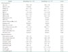
Table 1 shows the baseline clinical and laboratory parameters between two groups. Adverse remodeling occurred in 53 patients (25.5%) out of total of 208 patients. There were no significant differences in demographic factors between two groups. Of cardiac specific enzymes, only CK-MB was statistically different between the two groups. In case of PCI, there was no statistical difference in door to balloon time between two groups.
Echocardiographic characteristics
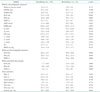
There were no significant differences follow up interval between two groups. EDV, end-systolic volume (ESV), DT, and GLS were found to be significantly different (p-value < 0.05) between two groups (Table 2). However, LVEF, GCS, and torsional parameters showed no significant association with LV adverse remodeling.
Predictors of adverse remodeling
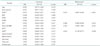
Table 3 shows the univariate and multivariate binary logistics analysis for adverse remodeling with respect to independent variables. Note that GCS, net twist, twist rate and untwist rate were not significant at both t-test and univariate analysis. Multivariate analysis was done with logistic regression with forward deletion. In univariate analysis, EDV [hazard ratio (HR): 0.953, 95% confidence interval (CI): 0.936–0.971, p< 0.001], ESV (HR: 0.968, 95% CI: 0.946–0.991, p = 0.006), DT (HR: 0.991, 95% CI: 0.984–0.999, p = 0.027), CK-MB (HR: 1.002, 95% CI: 1.001–1.004, p = 0.010), and GLS (HR: 0.884, 95% CI: 0.800–0.976,p = 0.015) were associated with remodeling. The variables that showed p-value of less than 0.100 in univariate analysis were used in the multivariate model; EDV, DT, peak CK-MB value, and GLS. Though ESV had p-value less than 0.1, it was excluded in multivariate analysis for its co-linearity with EDV. In multivariate binary logistics analysis, EDV (HR: 0.922, 95% CI: 0.897–0.948, p< 0.001), GLS (HR: 0.842, 95% CI: 0.728–0.974, p = 0.020), DT (HR: 0.989, 95% CI: 0.980–0.998, p = 0.023), and CK-MB (HR: 1.003, 95% CI: 1.000–1.005, p = 0.033) independently predicted LV adverse remodeling. However, GCS, net twist, and twist or untwist rate were not associated with remodeling.
Reproducibility
Variability in the measurement of strain was evaluated in 20 randomly selected patients. For intra-observer variability, the same observer (MJ Cha) measured strain for each of the selected patients again 15 days later. The correlation of intra-observer variability for GLS, GCS, and net torsion were 0.90, 0.83, and 0.78, respectively. For the inter-observer variability, a second independent observer (HM Na) repeated the analysis. The correlation of inter-observer variability for GLS, GCS, and net torsion were 0.84, 0.79, and 0.73, respectively.
Discussion
There has been extensive research about GLS, GCS and torsion and they were proposed as predictors for remodeling, however, most of them had limitation of small patient number.19)20)21)22)23) Some studies demonstrated that GLS as a powerful prognosticator for remodeling,19)20)23) while other studies claimed GCS17) or torsion22) is the best prognosticator. In this study, GLS, not GCS or torsion, predicted cardiac remodeling, in speckle tracking analysis. Beside of GLS, DT, baseline LV volume (ESV and EDV), and cardiac enzyme (peak CK-MB value) significantly predicted cardiac remodeling.
Park et al.19) demonstrated that GLS predicts remodeling. Although remodeling was defined by > 15% increase in EDV and they targeted only anterior wall infarct with patient population of fifty, their result was consistent with our study.
Lacalzada et al.20) also illustrated that GLS predicts remodeling in STEMI with ninety-seven low risk, patient group. This is consistent with our result even though only 20 out of 97 patients developed remodeling in that study. It is questionable that only GLS proved to be independent predictors among many powerful candidate variables such as cardiac enzyme, EDV, DM, status or LVEF in that study. This study proved EDV, DT, and CK-MB as additional significant factors with a population size over two times larger.
However, not all the studies were in agreement with our study. Previous studies focused on mostly high risk patients. We targeted low risk patients excluding patients older than 85-years-old, with atrial fibrillation, had to undergo CABG after CAG, and needed IABP or ECMO.
Unlike our study, Hung et al.17) demonstrated that only GCS rate independently predicted remodeling after high risk myocardial infarction, though both GLS and GCS predicted adverse outcome. In our study, GCS was not a predictor. In that study, of total patients, 50% had heart failure, 20% had LVEF of less than 35% and 30% had both, so that we may assume that those patients group are in very high risk for cardiac event. Furthermore, the mean follow-up period was more than 20 months. In our study, we excluded high risk patients; more than 90% of patients showed Killip class I and mean EF was more than 50%. Circumferential strain has been reported to be relevant to adverse clinical outcome in more advanced patients group.24)25) Longitudinal strain is more sensitive to early cardiac attack and circumferential strain may rather be preserved, initially.26)27) This is because subendocardium is more vulnerable to ischemia. Because longitudinal fibers are in subsubendocardial region and circumferential fibers are in the midwall region of the heart, the longitudinal fibers are more susceptible to ischemia. Thus, it leads to cardiac long-axis systolic dysfunction which can be reflected by impaired GLS. The shift from subendocardial dysfunction to subepicardial dysfunction is illustrated in previous study.25) Consequently, it seems that GLS may be early marker for cardiac dysfunction and GCS may be impaired in more advanced patients with decompensated heart. That is why GCS is not a predictor for remodeling in this study. This hypothesis should be further verified and also, prognosticators for this underlying irreversible process should be investigated.
Nucifora et al.22) reported that torsional strain independently predicted remodeling unlike to ours. The study population and those who reached the end point was relatively small in that study. The baseline characteristics of total patient were also different from this study. They had lower mean twist value (12.7° vs. 15.8° in our study) and lower EF (48% vs. 52%), which means that their study population had more advanced state than our study population. Another study showed that LV twist is related to infarct transmurality and is independently associated with LV remodeling after AMI.28) In that study, baseline EF and infarct related artery (left anterior descending artery territory) were not significantly different from our study. LV twist for its role in remodeling is still an area of unknown. Although it is well known that torsion is a key factor for systolic and diastolic function in cardiac mechanics, we have yet much to discover its role in cardiac remodeling.
Contrary to other studies, patients without LV remodeling had larger LV volume than with LV remodeling group. We do not know the exact reasons. However, we suggest possible explanations; 1) we included the patients with successfully reperfused by either PCI or thrombolysis, and therefore, LV volume was not larger than normal population in Korea.29) This may be related with more chances to get reverse remodeling in patients with larger LV volume. 2) Strict medication could be another cause of reverse remodeling. Core measure of Korean Health Insurance Review and Assessment Service forced to treat dual antiplatelet therapy, statins, beta-blockers, and renin- angiotensin system blockade in all STEMI patients, which was related that patients who had larger LV volume had more chances to reverse remodeling.
Limitations
We should consider some limitations of the study. It should be taken into account that it was a retrospective analysis, leading us to consider a selection bias. This study result cannot apply to all AMI group because we targeted only low risk STEMI patients. Considering that the echocardiography was done 24 hours after revascularization, it can be assumed that myocardial stunning might affect initial echocardiographic data. Thus, it might be different from the echocardiographic data after myocardial recovery. Although GLS had a prognostic power to predict remodeling in STEMI, area under curve for remodeling was only 0.62 (95% CI: 0.53–0.71). The best cutoff value does not have a high sensitivity and specificity for remodeling.
Although the quality of 2D speckle tracking imaging was quite clear, higher resolution for cardiac border is still needed. Also automated speckle tracking analysis can be possible through dedicated software. Furthermore, the border tracing needs manual work, hence intra- and inter-observer error was unavoidable. The development of speckle tracking is rapidly progressed. 3D speckle tracking may overcome many drawbacks of 2D strain in the near future.30)
 XML Download