Journal List > J Cardiovasc Ultrasound > v.24(2) > 1018421

See the article "".
Ryu: Normal Reference Values for Doppler Echocardiography: Influences of Ageing, Gender and Ethnicity
REFER TO THE PAGE [Related article:] 144-152
Echocardiography is now considered a standard diagnostic tool that enables the noninvasive quantification of cardiac chamber size, ventricular mass, and systolic function in various clinical situations. Technological progress in Doppler echocardiography has enabled the provision of hemodynamic information and the assessment of valvular heart disease and ventricular diastolic function.1)
When interpreting the results of medical tests, comparisons are usually made with a reference range that defines the values seen in health or considered desirable for health, and the effectiveness of any diagnostic test depends on its ability to accurately detect abnormalities. Echocardiography is no exception and it is essential to establish 'normal' reference values for echocardiography.2)
The population targeted and method used to collect the data are crucial for defining reference values. However, current Doppler echocardiography reference values are derived mainly from North American and European population studies with wide heterogeneity of their inclusion and exclusion criteria and may not be applicable to other populations.3) Reported normal reference values differ according to age, gender, and ethnicity.4)5)6) Several studies have examined these differences. However, the EchoNoRMAL study and an echocardiographic study of Latinos focused mainly on cardiac chamber size,7)8) while Okura et al.9) could not completely exclude subclinical conditions such as diabetes and renal failure that might have changes left ventricular (LV) diastolic parameters.
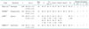
With increased awareness of the importance of accounting for age, gender, and ethnicity, several studies have obtained normal reference ranges for Doppler data for specific healthy populations,10)11)12)13) as listed in Table 1.
The Normal Echocardiographic Measurements in a Korean Population (NORMAL) study is the first Korean multicenter study providing reference values for the most useful Doppler parameters according to age and gender using conventional echocardiographic approaches.14) The NORMAL study ran from January 2011 to March 2014 and included 1003 normal adults (age 20–79 years) who had no significant cardiac disorders or clinical illnesses that might have affected cardiac structure and function, such as hypertension and diabetes. A previous study of normal echocardiographic reference values did not include tissue Doppler imaging (TDI) variables or provide sex-specific reference values.15)
The NORMAL Doppler study showed that normal ageing is associated with a number of changes in the heart and vascular system. Briefly, the mitral early diastolic inflow velocity (E), late diastolic inflow velocity (A), and E/A ratio were higher in women than in men. There were also considerable differences in the septal and lateral mitral annular velocities, and every TDI variable measured from the lateral annulus was greater compared to the values measured from the septal annulus. However, there were no significant differences in the septal and lateral early diastolic annular velocities (e') between men and women. The septal E/e' value in men was lower than in women.
The mitral E/A ratio, e' velocity, and E/e', which are representative variables used to evaluate diastolic dysfunction, decrease with age in both men and women. These results have been consistent in studies of European, Japanese, and Chinese populations and confirm that age reference values should be considered when analyzing diastolic function.10)11)12)13)16) These findings might partially explain why elderly people are likely to develop heart failure (HF) with a preserved ejection fraction and higher filling pressure.17)
However, in addition to the differences in the E/A ratio, e', and E/e' ratio between men and women among these studies, the changes with age were also not identical. This might be due to ethnicity, relatively small sample sizes, or the age and gender distributions of the study populations.
Ethnicity is an important determinant of the cardiovascular adaptation of systolic and diastolic function. Racial and gender differences in large-artery structure and function, endothelial function, the renin-angiotensin system, and levels of vasoactive cytokines may partially explain the differences.18) Also, in the studies mentioned above,10)11)12)13) the study populations ranged from 449 to 1394 and the proportion of males ranged from 44% to 55%. The numbers of participants in the various age groups were different.
As mentioned above, values of the mitral inflow E, E/A, and E/e' in men were significantly lower than in women, which is consistent with studies in Norwegian and Japanese populations.10)12) This suggests the need for gender-specific reference values in clinical practice. In women, HF is associated more with LV diastolic dysfunction than in men, whereas LV systolic dysfunction is the predominant cause of HF in men.19) Daimon et al.16) reported that for subjects < 50 years, women had a significantly greater E, E/A ratio, and e' than men, while these parameters were similar between the sexes in subjects > 50 years. Menopause usually occurs at the age of approximately 50 years, and the rapid changes in LV diastolic parameters in women over 50 seem to be consistent with postmenopausal status. The effects of estrogen and postmenopausal status on smooth muscle proliferation20) and vascular function21) may play a role in the gender-based differences in Doppler echocardiographic parameters.
In this study, patients with significant hypertension and diabetes were excluded based on the medical histories obtained from the study subjects.14) Therefore, patients with subclinical hypertension22) and diabetes mellitus,23) which are associated with diastolic HF, might be included in this study. This could partially contribute to the difference with previous findings.10)11)12)13)
In conclusion, heightened awareness of the importance of normal reference values according to ethnicity, age, and gender has emphasized the need for age- and gender-specific Doppler reference values for specific populations. In the future, high-quality comparative studies between different ethnicities and studies of the pathophysiologic effects of ethnicity, aging, and sex on Doppler echocardiographic parameters are needed for the correct diagnosis and management of systolic and diastolic dysfunction.

Figures and Tables

Table 1

Studies that provided normal reference values for variables from Doppler and TDI according to sex and age groups in specific ethnic group

jcu-24-112-i001

NORRE: Normal Reference Ranges for Echocardiography, JAMP: Japanese Normal Values for Echocardiographic Measurements Project, EMINCA: Echocardiographic Measurements in Normal Chinese Adults, TDI: tissue Doppler imaging, E: early diastolic inflow velocity, A: late diastolic inflow velocity, s': systolic annular velocity, e': early diastolic annular velocity, a': late diastolic annular velocity

Notes

Editorials published in the Journal of Cardiovascular Ultrasound do not necessarily represent the views of JCU or the Korean Society of Echocardiography.

References

1. Ommen SR, Nishimura RA, Appleton CP, Miller FA, Oh JK, Redfield MM, Tajik AJ. Clinical utility of Doppler echocardiography and tissue Doppler imaging in the estimation of left ventricular filling pressures: a comparative simultaneous Doppler-catheterization study. Circulation. 2000; 102:1788–1794.
2. Cosyns B, Garbi M, Separovic J, Pasquet A, Lancellotti P. Education Committee of the European Association of Cardiovascular Imaging Association (EACVI). Update of the echocardiography core syllabus of the European Association of Cardiovascular Imaging (EACVI). Eur Heart J Cardiovasc Imaging. 2013; 14:837–839.
3. Poppe KK, Doughty RN, Whalley GA. Redefining normal reference ranges for echocardiography: a major new individual person data meta-analysis. Eur Heart J Cardiovasc Imaging. 2013; 14:347–348.
4. Pfaffenberger S, Bartko P, Graf A, Pernicka E, Babayev J, Lolic E, Bonderman D, Baumgartner H, Maurer G, Mascherbauer J. Size matters! Impact of age, sex, height, and weight on the normal heart size. Circ Cardiovasc Imaging. 2013; 6:1073–1079.
5. Chahal NS, Lim TK, Jain P, Chambers JC, Kooner JS, Senior R. Ethnicity-related differences in left ventricular function, structure and geometry: a population study of UK Indian Asian and European white subjects. Heart. 2010; 96:466–471.
6. Munagala VK, Jacobsen SJ, Mahoney DW, Rodeheffer RJ, Bailey KR, Redfield MM. Association of newer diastolic function parameters with age in healthy subjects: a population-based study. J Am Soc Echocardiogr. 2003; 16:1049–1056.
7. Echocardiographic Normal Ranges Meta-Analysis of the Left heart (EchoNoRMAL) Collaboration. A meta-analysis of echocardiographic measurements of the left heart for the development of normative reference ranges in a large international cohort: the EchoNoRMAL study. Eur Heart J Cardiovasc Imaging. 2014; 15:341–348.
8. Qureshi WT, Leigh JA, Swett K, Dharod A, Allison MA, Cai J, Gonzalez F 2nd, Hurwitz BE, Shah SJ, Desai AA, Spevack DM, Rodriguez CJ. Comparison of echocardiographic measures in a hispanic/latino population with the 2005 and 2015 American Society of Echocardiography Reference Limits (The Echocardiographic Study of Latinos). Circ Cardiovasc Imaging. 2016; 9:e003597.
9. Okura H, Takada Y, Yamabe A, Kubo T, Asawa K, Ozaki T, Yamagishi H, Toda I, Yoshiyama M, Yoshikawa J, Yoshida K. Age- and gender-specific changes in the left ventricular relaxation: a Doppler echocardiographic study in healthy individuals. Circ Cardiovasc Imaging. 2009; 2:41–46.
10. Dalen H, Thorstensen A, Vatten LJ, Aase SA, Stoylen A. Reference values and distribution of conventional echocardiographic Doppler measures and longitudinal tissue Doppler velocities in a population free from cardiovascular disease. Circ Cardiovasc Imaging. 2010; 3:614–622.
11. Caballero L, Kou S, Dulgheru R, Gonjilashvili N, Athanassopoulos GD, Barone D, Baroni M, Cardim N, Gomez de Diego JJ, Oliva MJ, Hagendorff A, Hristova K, Lopez T, Magne J, Martinez C, de la Morena G, Popescu BA, Penicka M, Ozyigit T, Rodrigo Carbonero JD, Salustri A, Van De Veire N, Von Bardeleben RS, Vinereanu D, Voigt JU, Zamorano JL, Bernard A, Donal E, Lang RM, Badano LP, Lancellotti P. Echocardiographic reference ranges for normal cardiac Doppler data: results from the NORRE Study. Eur Heart J Cardiovasc Imaging. 2015; 16:1031–1041.
12. Daimon M, Watanabe H, Abe Y, Hirata K, Hozumi T, Ishii K, Ito H, Iwakura K, Izumi C, Matsuzaki M, Minagoe S, Abe H, Murata K, Nakatani S, Negishi K, Yoshida K, Tanabe K, Tanaka N, Tokai K, Yoshikawa J. JAMP Study Investigators. Normal values of echocardiographic parameters in relation to age in a healthy Japanese population: the JAMP study. Circ J. 2008; 72:1859–1866.
13. Yao GH, Zhang M, Yin LX, Zhang C, Xu MJ, Deng Y, Liu Y, Deng YB, Ren WD, Li ZA, Tang H, Zhang QB, Mu YM, Fang LG, Zhang Y. Echocardiographic Measurements in Normal Chinese Adults (EMINCA) Study Investigators. Doppler echocardiographic measurements in normal Chinese adults (EMINCA): a prospective, nationwide, and multicentre study. Eur Heart J Cardiovasc Imaging. 2016; 17:512–522.
14. Choi JO, Shin MS, Kim MJ, Jung HO, Park JR, Sohn IS, Kim H, Park SM, Yoo NJ, Choi JH, Kim HK, Cho GY, Lee MR, Park JS, Shim CY, Kim DH, Shin DH, Shin GJ, Shin SH, Kim KH, Park JH, Lee SY, Kim WS, Park SW. Normal echocardiographic measurements in a Korean population study: part II. Doppler and tissue Doppler imaging. J Cardiovasc Ultrasound. 2016; 24:144–152.
15. Park SW. Multicenter trial for estimation of normal values of echocardiographic indices in Korea. Korean Circ J. 2000; 30:373–382.
16. Daimon M, Watanabe H, Abe Y, Hirata K, Hozumi T, Ishii K, Ito H, Iwakura K, Izumi C, Matsuzaki M, Minagoe S, Abe H, Murata K, Nakatani S, Negishi K, Yoshida K, Tanabe K, Tanaka N, Tokai K, Yoshikawa J. Japanese Normal Values for Echocardiographic Measurements Project (JAMP) Study Investigators. Gender differences in age-related changes in left and right ventricular geometries and functions. Echocardiography of a healthy subject group. Circ J. 2011; 75:2840–2846.
17. De Sutter J, De Backer J, Van de Veire N, Velghe A, De Buyzere M, Gillebert TC. Effects of age, gender, and left ventricular mass on septal mitral annulus velocity (E') and the ratio of transmitral early peak velocity to E' (E/E'). Am J Cardiol. 2005; 95:1020–1023.
18. Lacolley P, Challande P, Osborne-Pellegrin M, Regnault V. Genetics and pathophysiology of arterial stiffness. Cardiovasc Res. 2009; 81:637–648.
19. Masoudi FA, Havranek EP, Smith G, Fish RH, Steiner JF, Ordin DL, Krumholz HM. Gender, age, and heart failure with preserved left ventricular systolic function. J Am Coll Cardiol. 2003; 41:217–223.
20. Ling S, Dai A, Dilley RJ, Jones M, Simpson E, Komesaroff PA, Sudhir K. Endogenous estrogen deficiency reduces proliferation and enhances apoptosis-related death in vascular smooth muscle cells: insights from the aromatase-knockout mouse. Circulation. 2004; 109:537–543.
21. Reis SE, Gloth ST, Blumenthal RS, Resar JR, Zacur HA, Gerstenblith G, Brinker JA. Ethinyl estradiol acutely attenuates abnormal coronary vasomotor responses to acetylcholine in postmenopausal women. Circulation. 1994; 89:52–60.
22. Bursi F, Weston SA, Redfield MM, Jacobsen SJ, Pakhomov S, Nkomo VT, Meverden RA, Roger VL. Systolic and diastolic heart failure in the community. JAMA. 2006; 296:2209–2216.
23. Galderisi M. Diastolic dysfunction and diabetic cardiomyopathy: evaluation by Doppler echocardiography. J Am Coll Cardiol. 2006; 48:1548–1551.
TOOLS
Similar articles