

 PDF
PDF ePub
ePub Citation
 Print
Print


Introduction
Carotid intima-media thickness (CIMT), a surrogate marker of subclinical atherosclerosis, has been consistently linked to cardiovascular and cerebrovascular disease (CVD).1)2) CIMT measurement by ultrasonography is simple and safe, uses no radiation, and is an inexpensive test to indirectly examine the presence of coronary atherosclerosis.3) Noninvasive high frequency ultrasonography gives valid estimates of the total arterial wall thickness and media thickness (MT) in carotid arteries.4) The typical B-mode image of the arterial wall is defined as the "double line pattern," which demonstrates that the inner line is generated by the intimal surface.5)
Associations between cardiovascular risk factors and CIMT have been established across a wide spectrum of ages, including children and adults.6)7)8)9)10) In a previous study, intima thickness (IT) increased exponentially with age. However, the increment of IT with age was different from that of MT.4)11) In hypertensive patients, the increment of CIMT was known to be caused by increased MT.12) Furthermore, with advancing age and through the development of atherosclerosis, changes occurring in the thickness of the intima and media layers are distinct from each other, also manifesting independent changes in different types of arteries.13) Therefore, separate measurements of the individual intima and media layers in carotid arteries might be more appropriate than the commonly used method of estimating the combined intima-media thickness. However, the clinical significance of separate measurements of CIMT, which is the sum of the IT and MT, to assess the risk for CVD has not yet been fully established.
In this study, we report the clinical usefulness of separate measurements of CIMT to assess subclinical atherosclerosis.
Methods
Study population
A total of 3377 patients underwent B-mode ultrasound of carotid arteries and coronary angiography (CAG) in the Medical Department of St. Mary's Hospital from September 2003 to March 2009. Seventy-nine percent of patients underwent CAG because of chest pain. The remainder underwent angiography for evaluation of arrhythmia, heart failure, congenital heart disease, valvular heart disease, or other problems. A total of 1146 subjects (M:F = 616:530; mean age, 57.7 ± 12.1 years) who were diagnosed with normal coronary arteries were enrolled in this study. There was no industry involvement in the design, conduct, or analysis of the study. The Ethics Committee approved the use of clinical data for this study, and all patients provided written informed consent.
Measurement of CIMT
The carotid arteries were evaluated using high-resolution B-mode ultrasound with a 15-MHz linear array transducer (HP Sonos-5500; Philips, Bothell, WA, USA). Carotid arterial scanning was performed by two certified sonographers, who were blinded to all clinical information, in a dark, air-conditioned room. With the patients in the supine position with slight hyperextension and rotation of the neck to the contralateral side, measurements of the CIMT were obtained throughout 10-mm segments across the far wall of the left and right common carotid arteries at a point which was most proximal to the carotid bifurcation. To optimize the image quality, the depth control was fixed at 4 cm. Measurements were acquired at the end-diastole phase under electrocardiography gating, because systolic expansion of the lumen causes CIMT thinning.14) CIMT was defined as the distance between the luminal border of the intima and the outer border of the media. The IT, high-echogenic intimal thickening (HEIT) to be exact, was assessed by manually measuring the thickness from the leading edge to the far edge of the first and second echogenic lines, and the media layer was defined as the distance between the two brightest echoes (Fig. 1).15) The IT/MT ratio was defined as IT divided by MT. The average of the IT, MT, and CIMT values of all segments was calculated to determine the mean IT, MT, and CIMT per patient. We repeated the individual measurements six times.
Clinical and biochemical assessment
Blood specimens were collected after a 12 to 14-hour fast (8:00 p.m. to 9:30 a.m.) to reduce the influence of circadian variation. The detail measurement was described in our previous study.16) The estimated glomerular filtration rate was determined by the original modification of diet in renal disease equation.
Blood pressure measurement
Blood pressure was measured with a mercury sphygmomanometer [first and fifth phases of Koroktoff sounds taken as systolic blood pressure (SBP) and diastolic blood pressure, respectively] after the subjects had comfortably rested for 5 min in the sitting position. An average of three measurements was used in the analyses. The measurement of blood pressure was performed right before and after the examination of CIMT. Systemic hypertension (HTN) was defined as systolic pressure ≥ 140 mm Hg and/or diastolic pressure ≥ 90 mm Hg, based on more than three measurements or current use of antihypertensive drugs.
Coronary angiography
Coronary angiograms were performed via the femoral or radial approach according to the American College of Cardiology/American Heart Association (ACC/AHA) recommendations for CAG. The angiograms were analyzed separately from the echocardiographic results by two experienced cardiologists. Coronary lesions were assessed with multiple orthogonal views and visually evaluated for the morphologic features similar to those reported by the ACC/AHA. Significant coronary stenosis was defined as ≥ 50% narrowing of the luminal diameter in one or more major coronary arteries. Coronary artery disease was defined as the presence of at least one coronary vessel with significant stenosis.
Patients with a principal complaint of chest pain and normal coronary artery on CAG underwent the acetylcholine provocation test. For the provocation test of coronary artery spasm, intracoronary injection of acetylcholine was administered according to the method of Okumura et al.17)
Statistical analysis
Continuous variables are presented as mean ± SD, and categorical variables presented as absolute and relative frequencies (%). The risk factor for atherosclerosis of IT, MT, CIMT, and the IT/MT ratio was assessed with univariate analysis and after adjustment for age and sex was analyzed. Multivariate analysis was performed to determine the independent risk factors of MT, mean and maximal CIMT, and the IT/MT ratio. All reported p-values were two-sided, and p-values of less than 0.05 were considered to indicate statistical significance. Intraclass correlation coefficient (ICC) is a statistical method for measurement of intra- and interobserver reliability. Reliability was assessed by replicating measurements for 50 patients. ICC assesses the consistency of multiple measurements of the same quantity. In general, the ICC ranges from 0.00 (no agreement) to 1.00 (perfect agreement). Also, an ICC value of 0.7–0.8 indicates strong agreement, and an ICC value > 0.8 indicates excellent agreement. ICC measures the proportion of the variance that is attributed to different observers. A 95% confidence interval was estimated for each ICC to estimate the precision and the range of the correlation. All statistical analyses were conducted in SAS 9.1 statistical software (SAS Institute Inc., Cary, NC, USA).
Results
Clinical characteristics
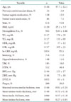
The baseline characteristics of the 1146 patients are shown in Table 1. The mean age of the study subjects was 57.7 ± 12.1 years, and 616 (53.8%) were men. All patients were diagnosed with normal coronary arteries confirmed by CAG. Most patients had atherosclerotic risk factors, such as HTN (n = 532, 46.4%), diabetes mellitus (DM) (n = 183, 16%), hypercholesterolemia (n = 148, 13%), and smoking (n = 216, 21%).
Inter- or intraobserver reliability of the separate measurements
To determine the reliability of the separate measurements of IT, MT, and CIMT, we use ICC, which is a statistical method for measurement of intra- and interobserver reliability. The inter- and intraobserver reliability for the separate measurements of IT, MT, and CIMT on the right side were 0.814 and 0.975, 0.900 and 0.993, and 0.965 and 0.978, respectively. The inter- and intraobserver reliability for the separate measurements of IT, MT, and CIMT on the left side were 0.760 and 0.977, 0.921 and 0.998, and 0.956 and 0.998, respectively
Atherosclerotic risk factors and separate measurements
In univariate logistic analysis, age, body mass index (BMI), triglycerides (TG), high-density lipoprotein cholesterol (HDL-C), high sensitivity C-reactive protein (hs-CRP), and HTN were associated with IT on the right side. Age, gender, BMI, hemoglobin A1c (HbA1c), renal function, cigarette-smoking status, HTN, and SBP were associated with MT and the IT/MT ratio on the right side. Age, BMI, HbA1c, renal function, cigarette-smoking status, DM, HTN, SBP, and left ventricle systolic function were associated with maximal and mean CIMT on the right side (Table 2). BMI was associated with IT on the left side. Age, renal function, history of HTN, and DM were associated with MT on the left side. Age, BMI, renal function, and HTN were associated with the IT/MT ratio on the left side. Age, HbA1c, renal function, HTN, and DM were associated with mean CIMT on the left side (Table 3).
In multivariate regression analysis, age, BMI, and history of HTN were associated with MT and the IT/MT ratio on the right side. Age, BMI, HbA1c, and history of HTN correlated with mean CIMT on the right side. Age and BMI were related to the IT/MT ratio on the left side. Age was associated with MT and CIMT on the left side (Table 4).
Discussion
This study shows that atherosclerotic risk factors, such as age, BMI, lipid profile, hs-CRP, and HTN seem to affect HEIT. In addition, the MT of the carotid artery is influenced by age, BMI, lipid profile, HTN, and SBP. Therefore, different atherosclerotic risk factors seem to affect the IT and MT of the carotid artery in different ways.
In the Bogalusa Heart Study8) and the Pathological Determinants of Atherosclerosis in Youth Study,18) non-invasive ultrasonographic evaluation of CIMT showed a strong relationship between traditional cardiovascular risk factors and early stages of vascular atherosclerosis in young adults.
Early vascular changes caused by atherosclerosis occur primarily in IT.4) Also, before development of atherosclerosis, diffuse intimal thickening (DIT) presents in arteries. The definition of DIT is intimal thickening spread out circumferentially and longitudinally in an arterial segment. The degree of DIT is evaluated based on the IT/MT ratio. The histological IT/MT ratio of the common carotid artery is about 0.1 in the 21- to 30-year-old age group and increases with age. Histologic IT/MT ratio of the coronary artery is larger than that of the common carotid artery.19) The HEIT and histologic IT were different. The HEIT value was larger than the histologically measured IT, possibly because the intima layer was damaged while harvesting and fixing the carotid artery.4) In an animal study, the IT represents about 15% on ultrasonography and 3.5% on histology. The HEIT is thicker than the histologic IT. The histologic IT/MT ratio and HEIT/MT ratio are different. However, the MT represents 70% of the total arterial wall on ultrasonography and 76% on histology. Ultrasonographic MT and histologic MT are similar. Therefore, total arterial wall thickness is mainly dependent on the MT.
Increased CIMT can also be seen with HTN due to medial hypertrophy or fibromuscular hyperplasia associated with aging.3)20) In a previous study, results suggested a differential response of the vasculature to systemic risk factors.10) One hundred subjects aged over 70 years with a diagnosis of CVD had a thinner MT and a thicker IT of the carotid arteries compared to healthy subjects.21)
However, the clinical significance of the separate measurements of CIMT in the patient with subclinical atherosclerosis has not yet been fully established. In this study, age had a close correlation with the thickness of all three arterial layers on both sides, even after the thickness was adjusted for other risk factors. Age, BMI, TG, HDL-C, hs-CRP, and HTN were statistically associated with IT. However, the difference between the value of IT with the risk factor and that without was 0.01 mm. Changes in the CIMT mainly depend on the MT. The MT was associated with age, BMI, and HTN in multivariate analysis. The IT/MT ratio in a patient with risk factors for atherosclerosis, such as age, obesity, HbA1c, cigarette-smoking status, and HTN was significantly smaller than the ratio in a patient without risk factors. In particular, in multivariate regression, the values of the IT/MT ratio on the right side in a patient with older age, obesity, and HTN were small. Therefore, the MT and the IT/MT ratio can give additional information about which risk factor may cause atherosclerosis.
Some limitations to our study deserve mention. First, we measured the CIMT in the far wall of both common carotid arteries, because the CIMT in the common carotid arteries is more reliable and less difficult to measure than in the carotid bifurcation or in the internal carotid arteries.7) However, measuring the CIMT in the common carotid, as well as the carotid bulb segments, is a more useful tool for detecting early stages of atherosclerosis and hypertensive changes in young adults.22) Second, we performed separate measurements of CIMT that were manually measured, and ultrasound imaging was an operator-dependent modality. To evaluate intra- and interobserver reliability, we used ICC, which is a statistical method for measurement of intra- and interobserver reliability. An ICC value of 0.7–0.8 indicates strong agreement, and an ICC value > 0.8 indicates excellent agreement. Third, the HEIT and the histologically measured IT were different, possibly because the intima layer was damaged while harvesting and fixing the carotid artery. The HEIT value was larger than the histologically measured IT. Nevertheless, the carotid HEIT correlated closely with the histologically measured IT.4)
As a large population study, this study indicates that separate measurements of the individual intima and media layers in carotid arteries are useful methods for assessing the effect of different atherosclerotic risk factors on the arterial wall, although the commonly used method of estimating the combined intima-media thickness did not evaluate the impact of the risk for atherosclerosis on IT and MT, respectively.


 XML Download