

 PDF
PDF ePub
ePub Citation
 Print
Print


Introduction
Several M-mode studies have noted abnormal septal motion in patients with constrictive pericarditis. In 1978, Candell-Riera et al.1) suggested that the "most outstanding echocardiographic sign of constriction was the recording of a brisk early diastolic movement of the interventricular septum (IVS), consisting of a rapid anterior displacement followed by a rebound toward the posterior left ventricular (LV) wall". Previous echocardiography Doppler studies have shown that ventricular inter-dependence, a result of fixed end-diastolic ventricular volumes causing coupling between the right and left ventricles is helpful in diagnosing constriction.2) As far as we are aware only one 2-dimensional (2-D) echocardiography study has described abnormal septal motion in detail in constriction, however, the specific septal movement was not described, and 2-D effects of inspiration on this motion were not examined.3) Magnetic resonance imaging (MRI) studies in constriction have also been performed but they lack details of the IVS movement, especially the specific effects of inspiratory manoeuvres. Cardiac catheterisation has been widely regarded as the "gold standard". The observation of the divergent increase in right ventricular (RV) pressure with inspiration associated with as decrease in LV pressure is widely acknowledged as a classic feature of constriction.4) Despite our current understanding, detailed echocardiography characterization of constrictive physiology (i.e., the identification of ventricular interdependence) and classification of severity based on these findings appear lacking in the literature.
Methods
In this single centre retrospective, observational study, we examined 2-D echocardiographic images of nine consecutive patients with constrictive pericarditis between 2005 and 2011 in our hospital. All patients underwent echocardiography using a Series 5 Vingmed cardiac ultrasound scanner. Standard views were obtained using a 2.5 MHz probe and images were retrieved from data stored on a large server. Images were reviewed by a single observer.
Six of the nine patients underwent MRI imaging. Assessment of ventricular coupling with real-time cine MRI was done on a series GE (General Electric, New York, USA) 1.5 Tesla Echospeed machine (software version 9.3i). Cardiac post processing was then performed on A GE AW work station. After scout views were done to localize the heart and determine the cardiac imaging plane positions. Cardiac and pericardial imaging morphology was studied using series of functional FIESTA images in 4 chamber and RV short axis plane. This was followed by a sequence of MR echo imaging in in-spiration and expiration.
A diagnosis of constrictive pericarditis is confirmed by cardiac catheterisation when there is a consistent increase in RV pressure during peak inspiration, at a time when the LV pressure is at its lowest.4)5) This respiratory discordance between LV and RV pressures is thought to be the most diagnostically reliable haemodynamic factor for distinguishing patients with constrictive pericardial disease with other disease entities.4)5) Five patients were investigated with cardiac catheterisation. Angiography was performed in a using simultaneous right and left heart catheterisation using high fidelity micromanometer catheters using shallow breathing and deep inspiration. Four patients underwent surgery for pericardiectomies.
Results
Baseline characteristics
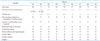
Table 1 summarizes clinical characteristics and investigations of these nine patients with constrictive pericarditis. The assessment of clinical severity of constriction was based on the presence of signs and symptoms. All patients experienced dyspnoea and had a positive Kussmaul's sign. Patients with moderate to severe constriction had pleural effusions and hepatosplenomegaly. Those with severe constriction had class IV New York Heart Association (NYHA) symptoms. Four patients underwent surgery and all were found to have definite constrictive pericarditis and their symptoms and echocardiography findings improved post-surgery. Patient P1 and P2 were in atrial fibrillation whereas other patients were in sinus rhythm. With all patients, there was at least 2 months delay in the diagnosis as most patients were initially treated for congestive heart failure. All patients had normal systolic LV function, and abnormal septal motion (in the absence of a bundle branch block). No patient had significant tricuspid regurgitation (TR) or pulmonary hypertension.
Echocardiographic findings
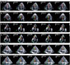
Resting and inspiration slow-motion 2-D echocardiographic analysis revealed abnormal septal motion in all patients with constrictive pericarditis. With normal, gentle respiration, patients in sinus rhythm and mild constriction (Patient P3) exhibited a very subtle, single wobble motion of the septum in diastole (Fig. 1A). Forced inspiration caused bowing of the septum into the LV cavity in diastole (Fig. 1B). This was noticed only in the first cardiac beat during the onset of inspiration. In patients with moderate constriction (Patient P2; figure not shown) this single wobble was more obvious and forced inspiration had a clear effect to bow the septum deeper into the LV cavity. With moderate to severe constriction (Patient P5, P6, and P7) the septum had an obvious double motion compared to the above patients during gentle respiration (Fig. 1C). This was characterized by a "bowing" or inversion of the septum into the LV cavity (Fig. 1C (ii)) in early diastole (after the T wave on the electrocardiography), followed by relaxation of the septum towards the central line (Fig. 1C (iii)). Following this there was further bowing of the septum into the LV cavity after atrial contraction (i.e., following the P wave) (Fig. 1C (iv)). The IVS then "resets" to the centerline prior to the next contraction (Figs. 1C (v) and 3). We have termed this motion "double wobble". Inspiration had little effect on this abnormal motion in the moderate to severe cases (Fig. 1D). Patients with severe constriction (P8 and P9) had only a single, prominent bowing motion of the septum into the LV cavity that lasted the duration of diastole ("pan-diastolic") (Fig. 1E). Inspiration had no effect on this motion (Fig. 1F). Two patients with atrial fibrillation and mild or mild to moderate constriction (Patient P1 and P2) demonstrated only a single motion into the LV cavity in diastole ("single wobble") with normal respiration and on inspiration.
MRI findings
Six of the nine patients underwent a cardiac MRI scan (Patient P1, P2, P3, P4, P7, and P9). Abnormal septal motion was seen in all six patients, in the pattern as described above for echocardiography. A single wobble motion was seen in patients with atrial fibrillation (Patient P1 and P2). A single wobble was identified in Patient P3 and P4 with mild and moderate constriction respectively. MRI was done in only one patients with moderate to severe constriction and MRI evidence of a double wobble motion was confirmed (Patient P7). Patient P9 had very severe constriction and the pan-diastolic motion was also confirmed with MRI.
Cardiac catheterisation findings
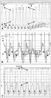
Five patients underwent cardiac catheterisation to assess for constrictive physiology (Patient P2, P4, P5, P7, and P9). All five were found to have elevated RV and LV diastolic pressures. Fig. 2A shows LV and RV pressures for P2 who had moderate constriction and evidence of a single wobble on echocardiography. It illustrates near equalization of pressures between the left and right ventricles during diastole (short downward pointing arrows). Tracings reveal slightly elevated LV diastolic pressures compared to the RV except during inspiration. With inspiration a divergent increase in RV and decrease in LV pressure was seen. This pattern is considered a diagnostic feature of pericardial constriction.2)4) The RV diastolic pressure is momentarily greater (first box; Fig. 2A). This is likely to explain the brief and noticeable movement of the IVS into the LV cavity during inspiration on echocardiography. We believe that the significant diuresis Patient P2 had undergone prior to catheterisation resulted in slightly reduced RV diastolic pressures compared to LV. We suspect that prior to diuresis (when the echocardiogram was taken) the RV diastolic pressure may have equalled or perhaps been greater than the LV to account for the wobble pattern seen on echocardiography. Fig. 2B illustrates findings for Patient P7 who also underwent significant diuresis by the time he had angiography. The first box in Fig. 2B shows RV diastolic pressure greater than the LV diastolic pressure after atrial contraction. We suspect that despite diuresis LV and RV diastolic pressures remained near equal at the time of catheterisation and supports our view that atrial contraction is likely responsible for the double wobble seen in these patients. Patient P9 who had severe constriction and pan-diastolic motion of the septum with bowing into the LV on echocardiography was found to have consistently higher RV than LV diastolic pressures despite significant diuresis (Fig. 2C). This explains the pan-diastolic motion of the septum into the LV cavity during diastole. With inspiration, the divergent increase in RV and decrease in LV pressure that are characteristic of constriction were again observed (broken arrow; Fig. 2C).
Discussion
Physiology of pericardial constriction
Pericardial constriction results from a rigid, noncompliant fibrous pericardium which creates a fixed end-diastolic ventricular volume so the outward expansion of the heart is impaired. As the total end-diastolic volume remains constant, any relative change in left or right ventricle volumes must be accommodated by septal movement, due to ventricular inter-dependence.
Abnormal septal motion with normal respiration in constrictive pericarditis
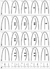
During normal gentle respiration, a single, subtle movement of the septum in diastole occurs in mild constriction (Patient P3) (Fig. 3A). This effect appears more pronounced in moderate cases. We found that patients with moderate and moderate to severe constriction (Patient P5, P6, and P7) had a double movement of the septum in diastole during normal respiration. The first deviation of the septum towards the LV cavity occurs with tricuspid valve opening in very early diastole (Fig. 3C). We believe that the increased volume in the right side results in deviation of the septum into the LV. The septum then straightens momentarily only to bow again into the LV cavity in mid to late diastole. The latter movement coincides with the onset of atrial systole and it is likely that increased emptying from the right atrium into the RV forces the septum towards the LV due to pericardial non-distensibility. The pan-diastolic bowing in severe constriction is due to the significantly increased right sided filling (compared to severely compromised left sided filling; Patient P8 and P9) (Fig. 3D) had end stage disease; both patients died. Two patients with constriction who were in atrial fibrillation had only a single early septal diastolic movement as seen in Patient P1 and P2, also suggesting that atrial contraction plays a role in the "second wobble" described above.
Important effect of forced inspiration
We found that forced inspiration caused prominent excursion of the septum into the LV which was most obvious during the first cardiac beat on early inspiration (Fig. 3B) in patients with mild or moderate constriction. This effect is likely due to enhancement of venous return due to the increased negative intra-thoracic pressure to the right side seen only momentarily as this pressure effect is brief in duration (duration of one contraction). Patient P5 who had more moderate rather than severe constriction (and a double wobble) did have increased excursion of the septum into the LV with inspiration in contrast to patients with more advanced disease (Patient P6 and P7) who did not. Inspiration had no effect on IVS motion in patients with severe constriction. This observation suggests that inspiration may be an important manoeuvre in not only identifying patients with mild constriction with only subtle wobble, but also in differentiating moderate cases from severe cases of constriction.
Abnormal septal motion also seen with severe tricuspid regurgitation
Patients with severe TR also have increased RV flow compared to LV and abnormal septal motion. We are unaware of any 2-D echocardiographic reports documenting the nature of IVS motion seen with TR but M-mode evidence does suggest abnormal motion.6) We examined 16 consecutive patients with severe TR and found that in all patients, subtle or significant pan-diastolic bowing of IVS into the LV cavity was seen and inspiration had no effect to augment or diminish the movement (data not shown). Clearly, the presence of congestive heart failure and severe TR will make the diagnosis of severe constriction more difficult to exclude in view of these findings.
Comparison of echocardiography, MRI and cardiac catheterisation
A previous MRI study described abnormal septal motion with constriction as early diastolic flattening in 17 of 21 patients. 7) Inversion (convex to the left) was seen in 2 of the 17 patients. Late diastolic septal inversion into the LV suggestive of a double wobble was not described, and changes with respiration were not undertaken. Another MRI study examined the influence of inspiration on septal motion patterns in patients with constriction. Inspiratory septal flattening or inversion was found in all 18 patients with constriction and 6 with inflammatory pericarditis, but it was not seen in 15 patients with restrictive physiology.8)9) Even though its accuracy in identifying abnormal septal motion, in our study we found MRI particularly challenging to perform in these patients due to difficulty in lying patients flat for long periods as many had significant dyspnoea. It is also difficult to coordinate the inspiration effort with image acquisition during MRI.
Cardiac catheterisation demonstrated an increase in RV pressure with inspiration with a concurrent decrease in LV pressure in all patients who underwent this manoeuvre. Catheterisation however is especially challenging in patients with severe constriction as they are unable to lie flat for long periods. By the time they undergo catheterisation they have usually undergone significant diuresis reducing cardiac pressures and hence the ability to identify ventricular inter-dependence, especially in mild cases. Echocardiography however is not constrained like MRI or catheterisation and appears sensitive especially in the acute setting when the patient first presents.
Conclusion
Our data suggest that 2-D echocardiography is a simple and readily available investigation that can identify pericardial constriction in symptomatic patients. Careful observation of the septal motion in addition to inspiration manoeuvres may help not only to identify constriction but also to classify its severity. Mild constriction causes a single, subtle wobble of the IVS which is more evident in moderate constriction. Moderate to severe constriction causes a double wobble, and severe constriction causes pan-diastolic movement. The limitations of this study include the retrospective and observational design using a small number of patients. We believe that these findings need to be validated with a larger prospective study.
 XML Download