

 PDF
PDF ePub
ePub Citation
 Print
Print


Introduction
Echocardiography is a useful diagnostic technique for detecting cardiac masses. Cardiac masses have various echocardiographic manifestations following etiology. Commonly observed intracardiac masses are tumors such as myxoma, metastatic tumor, or thrombi due to atrial fibrillation or systolic dysfunction, and vegetation due to infective endocarditis. Extrinsic lesion is mediastinal tumor, coronary and aortic aneurysm, and so on.
Diaphragmatic hernias include Morgagni hernia and hiatal hernia that are causes of cardiac mass lesion. However, they are relatively rare on routine echocardiographic procedures. Thus we can easily overlook or misdiagnose them. We present three cases of diaphragmatic hernia resembling cardiac mass on transthoracic echocardiography, which are subsequently identified hiatal hernia and Morgagni hernia.
Case
Case 1
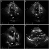
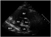
A 75-year-old female with hypertension was presented to our department because of dyspnea on exertion. On physical examination, blood pressure was 156/74 mm Hg. Crackle or murmur was not audible on auscultation. A 12-lead electrocardiogram showed normal sinus rhythm. Chest X-ray showed cardiomegaly and air-fluid level that suggest large hiatal hernia. A two-dimensional transthoracic echocardiogram showed huge extrinsic mass compress left atrium and filled with spontaneous echo contrast that has internal swirling flow (Fig. 1).
Case 2
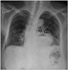
A 71-year-old female with prior history of hypertension and asthma presented neurosurgery department complaining of recently aggravated lower motor weakness and back pain. On physical examination, vital sign was stable. Crackle and murmur was not audible on auscultation. A 12-lead electrocardiogram showed normal sinus rhythm and left anterior fascicular block. Chest X-ray showed mild cardiomegaly, air-fluid level adjacent left cardiac boarder suggesting hiatal hernia, and blunting of left costophrenic angle suggesting left pleural effusion (Fig. 2). She underwent a two-dimensional transthoracic echocardiography for the preoperative evaluation.
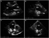
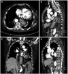
A two-dimensional transthoracic echocardiography showed huge extracardiac mass (3.8 × 3.5 cm) adjacent to the left heart compressing left atrium and mobile echogenic mass (3.4 × 2.8 cm) in right atrium (Fig. 3). The patient subsequently underwent chest computed tomography for differential diagnosis of mass lesion. Chest computed tomography showed large hiatal hernia in the posterior mediastinum (Fig. 4). After consulting a surgeon, the patient underwent an open hiatal hernia repair. Seven weeks later, a two-dimensional transthoracic echocardiography showed disappearance of preexisting mass adjacent to the left atrium and decreased echogenic mass in right atrium.
Case 3
A 28-year-old female without past medical history was presented to our department because of recently identified right pericardial mass like lesion on chest X-ray. But, she did not have any symptom. On physical examination, blood pressure was 125/75 mm Hg. Crackle and murmur was not audible on auscultation. A 12-lead electrocardiogram showed normal sinus rhythm. Chest X-ray showed opacity in right pericardial area suspected pericardial fat tissue. We performed a two-dimensional transthoracic echocardiogram and chest computed tomography to identify right pericardial opacity.
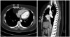
A two-dimensional transthoracic echocardiogram showed hypoechoic mass encroaching posterior aspect of both atria on parasternal short axis view (Fig. 5). Chest computed tomography showed prominent fat tissue in right cardiophrenic area with suspicious internally radiating vasculature that was suspected to be Morgagni's hernia (Fig. 6).
Discussion
Diaphragmatic hernia such as hiatal and Morgagni hernia can be mimic cardiac masses on transthoracic echocardiography. Previously, there was a report of hiatal hernia and Morgagni hernia that mimic pericardial lipoma.1) They only focused on differential diagnosis of hiatal hernia and echocardiographic findings wasn't concerned. For the rarity of these cases, it could be easily overlooked or misdiagnosed. So, we would like to report and discuss about that echocardiographic findings could give us useful information for the differential diagnosis of cardiac masses including hiatal hernia.
Hiatal hernia is characterized by the displacement of the gastroesophageal junction and part of the stomach into the mediastinum. Often asymptomatic, it may present wide spectrum of clinical manifestation from epigastric pain due to concomitant gastroesophageal reflux to postprandial syncope.2) It can be easily diagnosed with upper gastrointestinal barium examination or endoscopy. However, it can mimic a cardiac mass on transthoracic echocardiography. Echocardiographic feature that may suggest a hiatal hernia was first described by Nishimura et al.3) They reported that left atrial mass like lesion was in a maximal size when the left atrium was seen in a posterior plane, but smaller or absent as the left atrium was seen in more anterior planes. D'Cruz and Hancock4) reported additional findings. One is changes in the degree of encroachment of the hiatal hernia on the left atrium depending on respiratory cycle. Another is loss of the normal sharply defined sonolucency of the descending thoracic aorta due to superimposition of the hiatal hernia. The other is the visualization of swirling echodensities following the oral ingestion of carbonated beverage.4)
In case 1, transthoracic echocardiography showed spontaneous echo contrasts that had internal swirling flow without ingestion of carbonated beverage and this finding is helpful for diagnosing hiatal hernia. But we didn't undergo additional ingestion of carbonated beverage in case 2 and 3, because chest X-ray and computed tomography showed definite hiatal hernia in case 2 and computed tomography showed Morgagni hernia consisting of omental fat without stomach or bowel in case 3.
The first case showed typical echocardiographic findings hiatal hernia. At first, extrinsic mass that compressing left atrium is maximal size which was seen posterior plane. And it gradually decreased when it was seen anterior plane. The next, it showed internal swirling flow on parasternal ling axis view and apical 4-chamber view (Fig. 1). These findings were helpful for differential diagnosis of hiatal hernia without other imaging modalities.
Interestingly, although hiatal hernia is usually known to encroach on the left atrium, transthoracic echocardiography in the second case showed both atrial mass-like lesion separately because hiatal hernia was large enough to encroach both atria. So, we need to consider hiatal hernia as a differential diagnosis of extrinsic cardiac mass involving both atria.
Morgagni hernia is rarely reported diaphragmatic hernia due to defect of anterior diaphragm and first described by Giovanni Morgagni in 1769.5) Foramina of Morgagni is located immediately adjacent to the xiphoid process of the sternum and herniation occurred through it mostly right side of body.6) Mostly hernia sac contains transverse colon, omentum, liver, and, less frequently, small bowel or stomach. Morgagni hernia gets diagnosed late because it can be asymptomatic or present only ambiguous gastrointestinal or respiratory symptoms.7) In asymptomatic patient like this case, smooth supradiaphragmatic shadow of cardiac margin can be founded on routine chest X-ray. Nonetheless echocardiography has been shown to be useful, computed tomography is the most sensitive because it can give anatomical feature of the hernia and its complications such as strangulation.7) In our case, patient was asymptomatic and only had supradiaphragmatic shadow of right cardiophrenic angle on chest X-ray. Parasternal short axis view of transthoracic echocardiography showed encroachment of extrinsic mass lesion on the posterior aspect of both atria. Chest computed tomography showed prominent fat tissue in right cardiophrenic area and it contained radiating vasculature. These findings can be seen in Morgagni hernia that consists of omental fat and lipoma. The most important finding for differential diagnosis is that the presence of radiating fine linear or curvilinear densities indicates omental blood vessels and radiating vasculature indicates Morgagni hernia.8) Additionally, magnetic resonance imaging can be helpful. Since it readily provides images in the coronal and sagittal planes, it is better suited to recognize this diaphragmatic defect which lies predominantly in the axial plane.9)
In summary, diaphragmatic hernia such as hiatal and Morgagni hernia can be mimic cardiac masses on transthoracic echocardiography. As those were rare, we could easily overlook several specific echocardiographic findings which would help distinguish diaphragmatic hernia from other cardiac mass. So, we need to consider possibility of diaphragmatic hernia when we would find cardiac mass with specific echocardiographic features.
 XML Download