

 PDF
PDF ePub
ePub Citation
 Print
Print


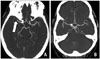
A 46-year-old Chinese man presented with left-sided weakness of sudden-onset for 145-minutes. His cardiovascular risk factors included hypertension and hypercholesterolemia. Neurological examination revealed left hemiplegia (Medical Research Council power grade 0/5) with National Institute of Health Stroke Scale (NIHSS) score of 16-points. An urgent non-contrast brain computed tomography (CT) scan was unremarkable while CT angiography revealed acute occlusion of the proximal right middle cerebral artery (MCA) (Fig. 1A). Treatment with intravenous tissue plasminogen activator (IV-tPA) was initiated at 175-minutes from symptom-onset. Continuous transcranial Doppler (TCD) monitoring of right MCA flow was performed using 2-MHz ultrasound transducer (Supplementary movie 1). No flow signals were noted in right MCA at the time of IV-tPA initiation. At 16-minutes, minimal antegrade flow (Fig. 2A) appeared in the right MCA that improved rapidly normal flow spectra within few seconds (Fig. 2B and C).1) Improvement in TCD flow signals was accompanied by rapid neurological recovery (NIHSS score decreased to 3-points at 20-minutes). He continued to improved and recovered considerably by day 2. CT angiography performed on day 2 revealed complete recanalization of the right MCA (Fig. 1B).
IV-tPA is the only approved therapeutic agent for achieving arterial recanalization in acute ischemic stroke. TCD is a noninvasive and bedside technique that can witness arterial recanalization in real-time during intravenous thrombolysis.2) Various randomized and observational studies provide an indication of the biological effect of TCD ultrasound in enhancing the effect of IV-tPA induced thrombolysis without any compromise on its safety regarding symptomatic intracranial hemorrhage.3) Our case demonstrates the rarely witnessed arterial recanalization of right MCA in real-time during intravenous thrombolysis and continuous TCD monitoring.

 XML Download