

 PDF
PDF ePub
ePub Citation
 Print
Print


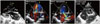
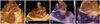
A 10-year-old child was referred to our department for the evaluation of dyspnea on effort of New York Heart Association functional class III. General examination was unremarkable. Cardiac examination revealed a normal first heart sound, loud second heart sound, no opening snap, a mid-diastolic and a pansystolic murmur at the apex and a grade III/IV ejection systolic murmur at the right second parasternal area. Two dimensional transthoracic echocardiography with color Doppler showed the presence of a partial supra mitral ring and a parachute mitral valve (PMV) leading to severe sub valvar mitral stenosis along with severe eccentric mitral regurgitation (Fig. 1A, B, and C, Supplementary movie 1and 2). There was also a subaortic membrane which resulted into severe subvalvar aortic stenosis (Fig. 1A). The aortic valve was quadricuspid (Fig. 1D) and there was no coarctation of aorta. Based upon these findings, a diagnosis of a partial form of Shone complex was made. Three dimensional (3D) transthoracic echocardiography clearly delineated the unifocal attachment of the PMV as well as the slit like opening in the inter chordal region that caused severe subvalvar mitral stenosis (Fig. 2A and B, Suppplementary movie 3). The near-circular morphology of the sub aortic membrane with a central opening was also evident on 3D echocardiography when viewed in the parasternal orientation (Fig. 2C, Supplementary movie 4) and from the left ventricular side (Fig. 2D). Patient is now planned for surgical intervention with possible repair of the mitral valve and resection of sub aortic membrane.
Shone et al.1) first described a complex congenital cardiac anomaly in 1963 which included PMV, supra mitral ring, sub aortic stenosis and coarctation of aorta. Partial forms of Shone complex are also described where only two or three out of four obstructive lesions are present. A good outcome is possible in patients with this rare anomaly if the surgical intervention is undertaken early before the onset of pulmonary hypertension.2) Precise anatomical imaging of the PMV as well as the sub aortic membrane is necessary before the surgical intervention. Our case demonstrates how cropping the 3D data set can give rise to unique imaging views for sub valvar structures like chordal attachments of the PMV as well as the near-circular morphology of the sub aortic membrane which were not evident on two dimensional echocardiography.
 XML Download