

 PDF
PDF ePub
ePub Citation
 Print
Print


Introduction
The definition of metabolic syndrome (MS) consists of at least three risk factors associated with type 2 diabetes mellitus and cardiovascular disease (CVD).1)2) In recent days, the prevalence of childhood MS has increased dramatically due to the increasing rate of childhood obesity.3)
However, research on this disease has been difficult because of lack of agreement on a definition of childhood MS. Until now, most definitions have been adapted from the Adult Treatment Panel III definition.4) In addition to this definition, the International Diabetes Federation released its definition of MS in children and adolescents in 2007 in order to measure the global prevalence of MS between nations.5)
Visceral obesity and insulin resistance may appear to be the important factors of the development of MS. Visceral fat accumulation is related with atherosclerosis and adverse cardiovascular consequences, as many pro-inflammatory cytokines are secreted from visceral adipose tissue.1)3)
MS has not been well studied on pediatric populations.6) Although, it is known that as the risk factors for MS increase, the occurrence also does. But, the underling pathogenesis of MS remains unclear.
Recently, tissue Doppler imaging (TDI) was reported to be appropriate for evaluating early changes in systolic and diastolic left ventricle (LV) function.7)8) Detection of myocardial deformation by TDI during the subclinical period in obese patients is important in clinical follow-up and in determining the prognosis.9)10)11)12)
Strain and strain rate imaging (SRI) have been proposed as a noninvasive echocardiographic technique to quantify regional myocardial function at rest and during acute and chronic ischemia.13)14) SRI is independent of cardiac rotational motion and a tethering effect.15)
There are several studies in adult with MS by strain and SRI. However, the results are controvertial.16)17)18)19) The efficacy of strain and SRI has not been determined in adolescents with MS. There are limited data on subclinical LV systolic dysfunction in overweight and morbid obese children.7)9)
The aim of this study was to investigate cardiac structural and functional alterations in adolescents with MS and to compare those with control subjects to screen for MS in asymptomatic adolescents and to evaluate the impact of MS on global LV function by TDI and SRI.
Methods
Study population
Ninety one healthy male adolescents aged between 14 and 15 participated in this study. They were divided into two groups based on the presence or absence of MS (MS group, non-MS group). Obesity was defined by body mass index (BMI) above the 95th percentile for age and sex as it is defined by Korean Centers of Disease Control and Prevention in 2007. People with endocrine dysfunction or other illnesses were excluded. We obtained informed consent from their guardians after explaining the procedure and purpose of the test thoroughly, and the study was approved by the Institutional Review Board of Ewha Womans University.
Diagnosis of metabolic syndrome
The diagnosis of MS was based on modified Cook's definitions (Table 1). In our study, the cut off value of fasting glucose was 100 mg/dL. MS was defined as having more than three components for their age and gender of the following metabolic abnormalities: abdominal obesity, [waist circumference (WC) ≥ 90 percentile], hypertriglyceridemia [serum triglyceride ≥ 110 (mg/dL)], low high density lipoprotein-cholesterol (HDL-C < 40 mg/dL), high fasting plasma glucose (fasting plasma glucose ≥ 100 mg/dL), and high blood pressure [≥ 90 percentile (mm Hg)].
Anthropometric measurement
We obtained anthropometric data of weight, height, WC, BMI, fat mass, and fat % from both groups. WC was measured with a tapeline at the mid-waist point between the lowest margin of the 12th rib and the mid portion of the superior iliac crest at minimal respiration. BMI was calculated by dividing the body weight in kilograms by height in meters. Fat mass and fat % were estimated by bioelectric impedance analysis (InBody 720, Biospace Co., Ltd., Seoul, Korea).
Blood pressure was measured using an automatic oscillometric method, Dinamap, Procare-200 (GE Medical System, Milwaukee, WI, USA) in a supine position after 10 minutes of adequate rest.
Laboratory measurement
Blood was drawn from all 91 adolescents, who had fasted for 14 hours prior to their blood sampling to determine the following parameters: blood glucose, total cholesterol (TC), HDL-C, low density lipoprotein-cholesterol (LDL-C), triglyceride (TG), aspartate aminotransferase, alanine aminotransferase (ALT). And, high sensitive C-reactive protein was also estimated.
Insulin resistance was measured by the homeostasis model assessment of insulin resistance (HOMA-IR), which was calculated by dividing the multiple of insulin (µU/mL) and serum glucose (mmol/L).
Echocardiographic parameters
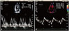
Echocardiography was performed using the IE33 machine (Philips Medical System, Andover, MA, USA) with an S5-1 transducer. Standard parasternal and apical views were acquired. Complete two-dimensional (2D) and M-mode echocardiogram, pulsed, color-flow Doppler, and TDI (Fig. 1) were obtained in the left lateral decubitus position. We measured the following LV parameters by M-mode echocardiography: interventricular septal wall thickness, posterior wall thickness, and LV end diastolic dimension at the chordae tendina level. The LV mass (LVM) and LV mass index (LVMI) were calculated. Ejection fraction (EF) was determined by using the biplane Simpson formula and fractional shortening was calculated using LV internal dimensions.
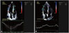
The diastolic function was assessed with pulsed Doppler mode from the apical window. Early diastolic (E), late atrial (A) peak velocities, E/A ratios, and deceleration time were performed using conventional pulsed wave Doppler echocardiography (Fig. 2).
To obtain longitudinal myocardial velocity, strain and strain rate with high quality, a narrow sector angle was used and image depth was adjusted to allow for a high frame rate (130-160 frames/s) with care taken to avoid angulations. The myocardial time-velocity and time-deformation curves were reconstructed off-line from color coded 2D tissue Doppler image loops. The peak systolic (s'), early diastolic (e'), and late diastolic longitudinal myocardial velocities (a') were measured at the basal septum from 4-chamber views (Fig. 2).
A single investigator who was blinded to the clinical data performed. Circumferential strain curves from the short-axis view (six segments: anteroseptal, anterior, lateral, posterior, inferior, and septal) and longitudinal strain curves from the apical four-chamber view (six segments: basal interventricular septum, basal anterolateral, medium interventricular septum, medium anterolateral, apical septum, apical lateral septum). Global strain was calculated as the average of the segments when six segments were measurable. Peak systolic strain rate was measured (Fig. 2).
Carotid intimal medial thickness
cIMT was measured with an ultrasonogram to which a 12 MHz linear transducer was installed (CX50 machine; Philips Medical System, Andover, MA, USA). cIMT by an experienced technician unaware of the group to which the subject patients were assigned. The subject patients were given the test in a lying position with their head slight turned. Within 1 cm from the junction of the right common carotid artery, the cIMT and vascular diameter of the vessel during the systolic and diastolic periods were measured. cIMT was defined as the distance between the two points which were observed as bright images on echocardiography.
Pulse wave velocity and ankle brachial index
Brachial-ankle pulse wave velocity (baPWV) and ankle brachial index were measured in a supine position using a volume-plethysmographic apparatus (Colin Co. Ltd., Komaki, Japan). The average of the left and right baPWVs in each subject was used as the PWV.
Statistical analyses
We performed all statistical analyses using Statistical Package for the Social Sciences (SPSS) (version 20, SPSS Inc., Chicago, IL, USA). Descriptive statistics were presented as means and standard deviations. The comparison of continuous variables was done using the Wilcoxon rank-sum test or one-way analysis of variance. A p-value less than 0.05 was considered as statistically significant.
Univariate analysis was performed to investigate the correlations between MS and other metabolic parameters.
Results
Prevalence of each component of MS
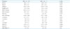
The most common symptoms of the criteria were HDL-C < 40 mg/dL (100%), fasting glucose (85.7%), TG ≥ 110 mg/dL (71.4%), followed by WC ≥ 90 percentile (42.9%), systolic blood pressure (SBP) or diastolic blood pressure ≥ 90 percentile mm Hg (42.9%) (Table 3).
Comparison of anthropometric data
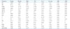
Weight, BMI, and WC were significantly higher in the MS group compared with the non-MS group (Table 4).
Comparison of biochemistry data
In the laboratory tests, glucose, insulin, HOMA-IR, and TG were significantly higher in the MS group compared with the non-MS group.
HDL-C was significantly lower in the MS group compared with the non-MS group (Table 5).
Comparison of echocardiographic data
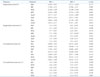
LVM was significantly higher in the MS group compared with the non-MS group (192.0 ± 73.0 g vs. 140.6 ± 30.2 g, p < 0.05, p < 0.05). LVMI were significantly higher in the MS group compared with the non-MS group (67.6 ± 22.3 g vs. 51.5 ± 10.4 g, p < 0.05) (Table 6).
e' was significantly lower in the MS group compared with the non-MS group (10.1 ± 2.9 cm/sec vs. 11.7 ± 2.1 cm/sec, p < 0.05). s' was significantly lower in the MS group compared with the non-MS group (6.6 ± 1.0 cm/sec vs. 7.8 ± 1.2 cm/sec, p < 0.05). Global longitudinal strain (GLS) was significantly lower in the MS group compared with the non-MS group (-17.5 ± 2.0% vs. -19.5 ± 2.7%, p < 0.05) (Table 6).
There was a significant reduction in longitudinal strain at the LV medium anterolateral septum (-21.68 ± 7.00% vs. -13.28 ± 5.56%, p < 0.05), the medium interventricular septum (-23.82 ± 4.89% vs. -17.14 ± 3.18%, p < 0.05) and the apical anterolateral longitudinal septum (-16.77 ± 5.27% vs. -12.43 ± 4.86%, p < 0.05) (Table 7).
The longitudinal strain rate (LSR) was significantly lower in the medium interventricular septum in the MS group compared with the non-MS group (1.71 ± 0.52 s-1 vs. 1.17 ± 0.64 s-1, p < 0.05) (Table 7).
Intimal medial thickness and pulse wave velocity
cIMT and pulse wave velocity were not significantly different between two groups (Table 8). The compliance and distensibility of the carotid artery were calculated. There was not any significant difference between two groups. Data is not shown.
Linear correlation between cardiac parameters and other anthromometric data
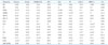
LVMI was significantly correlated with BMI (r = 0.39, p < 0.05), fat mass (r = 0.36, p < 0.05), SBP (r = 0.34, p < 0.05), fat % (r = 0.33, p < 0.05), WC (r = 0.32, p < 0.05), and weight (r = 0.31, p < 0.05).
e' was significantly correlated with BMI (r = -0.25, p < 0.05), fat % (r = -0.22, p < 0.05), and fat mass (r = -0.21, p < 0.05).
Basal anterolateral LSR was significantly correlated with WC (r = 0.25, p < 0.05), fat mass (r = 0.24, p < 0.05), BMI (r = 0.22, p < 0.05), fat % (r = 0.21, p < 0.05), and weight (r = 0.26, p < 0.05) (Table 9).
Linear correlation between cardiac parameters and blood chemistry data
LVMI was significantly correlated with ALT (r = 0.34, p < 0.05), TC (r = 0.24, p < 0.05), and LDL-C (r = 0.20, p < 0.05). e' was significantly correlated with HDL-C (r = 0.22, p < 0.05).
s' was significantly correlated with LDL-C (r = 0.23, p < 0.05). GLS was significantly correlated with insulin (r = 0.23, p < 0.05), HOMA-IR (r = 0.22, p < 0.05), and TG (r = 0.2, p < 0.05).
Global circumferential strain (GCS) was significantly correlated with glucose (r = 0.22, p < 0.05) and TC (r = 0.21, p < 0.05) (Table 10).
Linear correlation between insulin resistance and other parameters
HOMA-IR was significantly correlated with GLS (r = 0.22, p < 0.05) (Table 10).
Discussion
In our study, adolescents with MS showed subclinical LV systolic and diastolic dysfunction by TDI and SRI. LVM and LVMI were significantly higher in the MS group compared with the non-MS group. e', s' velocity and GLS were significantly lower in the MS group compared with the non-MS group. There was a significant reduction in strain at the LV medium anterolateral septum, the medium interventricular septum and the apical anterolateral longitudinal septum. The LSR was significantly lower in the medium interventricular septum in the MS group compared with the non-MS group.
Utility of TDI and SRI have not been determined in pediatric patients with MS.
Previous adult studies have investigated the LV functions in patients with MS, but there is not any consensus.16)17)18)19) Grandi et al.17) have described that only LV diastolic function is reduced in MS, although LV systolic function is normal. Masugata et al.18) also have reported that cardiac diastolic function was impaired in patients with MS even if they have neither LV hypertrophy nor systolic dysfunction. On the other hand, Wong et al.19) have found that MS is correlated with both LV systolic and diastolic dysfunction in subjects with significant risk factors but not with CVD. Those studies have provided valuable information about LV function in MS.
However, there are some limitations to compare our data with those studies, because our study was performed in adolescents, the other studies were done on adult age patients.
Diagnostic methods used to assess cardiac function are different between several studies. Gong et al.16) found that regional LV systolic and diastolic function were impaired in patients with MS by using strain and SRI.
The impairment was more serious in patients with four risk factors than in patients with three risk factors. This suggests that different components of MS may have synergistic effects on LV functions.
In our study, ventricular systolic and diastolic function were significantly lower in the MS group with more than 3 risk factors compared with non-MS group with less than 3 risk factors.
These results were in accordance with the previous studies in adult groups.16)
Although the cause of altered LV function in MS is not fully understood, the components of MS may produce latent cardiac structural and functional disorders. MS is also related to LV hypertrophy and LV diastolic dysfunction.20)
In our studies, e' velocity showed significant negative correlation with BMI, fat %, fat mass. e' was significantly correlated with HDL-C.
GLS was significantly correlated with insulin, HOMA-IR and TG. GCS was significantly correlated with glucose and TC. Basal anterolateral LSR was significantly correlated with weight, WC, fat mass, BMI, and fat %. LVM was significantly correlated with ALT and glucose.
Obesity is associated with increased metabolic demand because of greater adipose tissue, larger blood overload and increased preload to the heart. Also, vascular alterations of arterial stiffness and resistance increase afterload to the heart. Obesity can cause cardiac hypertrophy in systolic and diastolic cardiac dysfunction.21)
Obesity affects myocardial metabolism. Alterations in myocardial substrate metabolism have been related with reduced myocardial contractile dysfunction in insulin resistant patients and diabetes mellitus.19) LV abnormalities seem to be greater in visceral rather than in subcutaneous obesity.22) The cause of this myocardial dysfunction remains unclear, although chronic volume overload, insulin resistance, autonomic changes and metabolic abnormality have already been studied.
BMI, insulin resistance and duration of obesity are associated with cardiac dysfunction.23)
Increased adiposity is associated with insulin resistance because of the release of free-fatty acid, as well as actions of adipokines. Lipotoxicity can also contribute to cardiac myocyte apoptosis.19) Other possible mediators include the hormonal and cytokine release associated with obesity. Adiponectin is reduced in obesity, and this may induce insulin resistance.24)
Our results suggest that obesity contributes to the myocardial changes. Interestingly, we found that HOMA-IR was significantly positively correlated with GLS (r = 0.22). Hyperinsulinemia and insulin resistance have been known to cause an altered collagen/muscular ratio.25) Studies have demonstrated an accumulation of myocardial collagen is related to interstitial and perivascular fibrosis, both of which is associated with LV early diastolic dysfunction.26)
In our study, LV hypertrophy, which was assessed by the LVMI was significantly different between patients with MS and those in control adolescents. LVMI was significantly correlated with BMI (r = 0.39), fat mass (r = 0.36), SBP (r = 0.34), fat % (r = 0.33), and WC (r = 0.32). LVMI was significantly correlated with ALT (r = 0.34), TC (r = 0.24), and LDL-C (r = 0.20).
Both eccentric and concentric hypertrophy have been noted in adults with obesity and are impacted by the degree and duration of the obesity.27) Altered cardiac morphology may be a precursor to impaired cardiac function. Studies on children and adolescents with obesity have reported changes in cardiac mechanics including diastolic dysfunction28) and systolic dysfunction at rest29)30) and during exercise.31)
Currently, it is uncertain how cardiac dysfunction will progress over time, and how cardiac dysfunction correlates to outcomes in adulthood. In adults, even with cardiac enlargement and systolic dysfunction, several studies have revealed that LVEF is normal in many obese subjects.32) Although the EF is normal, myocardial contractile abnormalities may still exist, and are more susceptible to measures by TDI and SRI analyses.33)
TDI is used to estimate LV filling pressures in obese subjects. The ratio of early diastolic filling to mitral annular tissue velocity (E/E1) is a well-established index of pulmonary capillary wedge pressure strain.34)
In our study, we performed the measurements to keep the angle between the ultrasound beam and the LV longitudinal axis as small as possible to eliminate errors and the data were averaged using a compound function to decrease noise by 2D speckle tracking imaging.
The traditional method of deriving regional systolic strain and strain rate from TDI is, however, limited by angle dependency.
We found that regional strain and strain rate reduction in the MS group with normal LVEF. The use of TDI and SRI analysis could detect pre-clinical changes in LV regional systolic and diastolic dysfunction before conventional changes in EF.35)
Previous studies of MS in children and adolescents, gave evidence on the clustering of risk factors for CVD.36) Although there hasn't been any research that has shown the impact of MS on heart disease in childhood, some autopsy studies in childhood have shown that the factors of obesity, high blood pressure, high TG, and low HDL-C are related to the early stage of coronary atherosclerosis.19)22) Early identification of the clustering of risk factors in childhood is important in targeting efforts for chronic disease prevention.
There are several limitations to this study. This study only analyzed male subjects. There is also a lack of comprehensive assessment according to diverse growth and development because the age was confined to ages between 14 and 15 years old. Another limitation of this study includes the accuracy and reproducibility of measuring the parameters using echocardiography.
Further research on a larger adolescent population is needed, because there is no standard cut-points for parameters of MS. The longitudinal model design from adolescence to adulthood is needed to explore the complexity of MS risk factors. Since the time course of an adolescent to obesity and clinical disease is short, there is a lack of difference between the MS group and the non-MS group. The prospective and longitudinal studies may offer good opportunities in pediatric cardiovascular research.
In conclusion, in the present study, we have shown the presence of impaired global left ventricular function in patients with MS compared with control subjects without MS even if they have normal LVEF. This finding emphasizes the importance of early diagnosis and management of MS to prevent the progression of ventricular dysfunction to overt structural and symptomatic cardiac disease.
Early identification of regional and global LV functions by TDI and SRI in MS risk group may help to stratify risk and guide therapy. Because the components of the MS tended to be highly correlated with each other, it would be difficult to detect completely separate effects of each component. Strain and SRI would be a sensitive and feasible method to detect subclinical abnormalities in those populations.


 XML Download