

 PDF
PDF ePub
ePub Citation
 Print
Print


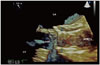
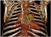
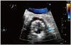
Valve-in-valve trans-cutaneous aortic valve implantation (TAVI) is now feasible and in a recent multi-centre study showed performed with high technical success rates, acceptable post-procedural valvular function, and excellent functional improvement.1) Multi-modality cardiac imaging in such patient's is slowly becoming routine and it is becoming pivotal for physicians to recognize structural and anatomical changes present in such patients. A 70-year-old gentleman was admitted to our unit with pyrexia and suspected infective endocarditis. He had previously undergone bio-prosthetic aortic valve implantation in 2004 for aortic stenosis. When he re-presented with heart failure in 2012 due to re-stenosis of the aortic valve bio-prosthesis, he was deemed not suitable for re-do surgery because of high-risk involved. Thus, he had TAVI using Medtronic CoreValve. Three-dimensional transoesophageal echocardiogram (3D TEE) and multi-slice contrast enhanced computed tomography (CT) scan were performed to look for the source of infection. 3D TEE clearly showed the grove of CoreValve in aortic sinus and that it was well seated (Fig. 1). Position and location of the CoreValve was also confirmed on CT thorax with 3D vascular multi-slice reconstruction (Fig. 2). Two-dimensional short-axis colour Doppler views confirmed patent left main stem and location of the strut in relation to it (Fig. 3). The highlight of this peculiar case is that the CoreValve can be recognised by 3D TEE and also CT. However, it is not possible to distinguish the remnant leaflets of the previous bio-prosthesis. This is important to bear in mind when performing and reporting TEE and CT in patients with aortic valve in valve systems. To best of our knowledge, this state-of-the-art 3D-volume rendered multi-modality imaging assessment of valve in valve TAVI has not been reported previously in literature.
Figures and Tables
Fig. 1
Three-dimensional transoesophageal view of Medtronic CoreValve (blue arrows) embedded in sinus of Valsalva, where bio-prosthetic aortic valve was previously implanted. In this systolic view the CoreValve cusps are open (red arrows). LA: left atrium, LV: left ventricle, LVOT: left ventricular outflow tract.
References
1. Eggebrecht H, Schäfer U, Treede H, Boekstegers P, Babin-Ebell J, Ferrari M, Möllmann H, Baumgartner H, Carrel T, Kahlert P, Lange P, Walther T, Erbel R, Mehta RH, Thielmann M. Valve-in-valve transcatheter aortic valve implantation for degenerated bioprosthetic heart valves. JACC Cardiovasc Interv. 2011; 4:1218–1227.

 XML Download