Journal List > J Cardiovasc Ultrasound > v.22(4) > 1018343

Moustafa, Alvarez, Connelly, Prieur, Patton, and Mookadam: Severe Form of Tetralogy of Fallot: Late Presentation
A 25-year-old man was evaluated due to progressive exercise intolerance and desaturation in the setting of previously documented Tetralogy of Fallot (TOF). He was diagnosed with TOF shortly after birth but his family declined surgery. Three years earlier, he had undergone left sided modified Blalock-Taussig shunt as a palliative procedure due to progressive shortness of breath and desaturation (O2 saturation between 80-85%). Initially, in the first few months after the procedure, he noticed slight improvement of his symptoms. However, he developed progressive exercise intolerance and desaturation which mandated complete repair. Physical examination was remarkable for desaturation (O2 saturation between 85-90%), clubbing and systolic ejection murmur (grade II-III/VI) over the pulmonic area. Electrocardiogram showed right axis deviation and right ventricular (RV) hypertrophy. Chest X-ray revealed dilated right side of the heart.
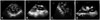
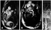
Transthoracic echocardiogram (TTE) was technically difficult still showed dilated hypertrophied RV with normal systolic function, malalignment ventricular septal defect (VSD) and overriding of the aorta (Supplementary movie 1 and 2). Transesophageal echocardiogram (TEE) was carried out for further delineation of the RV outflow tract (RVOT) which was hypoplastic with a long (~4 cm) segment of narrowing and significant infundibular narrowing (by hypertrophied muscle bundle). Peak gradient across the RVOT was approximately 35-40 mm Hg (Fig. 1, Supplementary movie 3 and 4). Cardiac magnetic resonance (CMR) was technically challenging due to patient's non-compliance with breathing instructions, yet confirmed the findings of TTE/TEE. Additionally, CMR uncovered hypoplastic proximal main pulmonary artery (PA), dilated left PA and mild proximal right PA stenosis with hypoplasia throughout the course (Fig. 2, Supplementary movie 5). The patient underwent successful complete repair with RV to PA conduit, resection of the native RVOT muscle bundles, arterioplasty of the right PA and VSD pericardial patch closure. Surgery was uneventful.
An amalgamated approach of echocardiogram and CMR offers a precise detailed assessment of the RV, RVOT, and PA, which is critical for decision making regarding surgical intervention in this unusual case of unrepaired TOF presenting late in adult life.1)2)

Figures and Tables

Fig. 1
A: Transesophageal echocardiogram (TEE) transgastric view showing dilated right ventricle with significant hypertrophy. B: TEE apical long axis view showing overriding of the ascending aorta with malalignment ventricular septal defect (arrow). C: TEE transgastric view showing hypoplastic right ventricular outflow tract (arrow) with infundibular stenosis. Pulmonic valve is shown (arrowhead). D: TEE modified mid esophageal view showing significant infundibular stenosis (arrow). AO: ascending aorta, LV: left ventricle, PA: pulmonary artery, RV: right ventricle.
jcu-22-228-g001
Fig. 2
A: Cardiac magnetic resonance (CMR) sagittal image showing overriding of the ascending aorta with malalignment ventricular septal defect (arrow). B: CMR sagittal image showing hypoplastic right ventricular outflow tract and proximal main pulmonary artery (arrow). C: CMR angiography showing hypoplastic right ventricular outflow tract, main pulmonary artery (arrow) and right pulmonary artery (RPA) with severely dilated left pulmonary artery (LPA). AO: ascending aorta, LV: left ventricle, PA: pulmonary artery.
jcu-22-228-g002

References

1. Nakamori S, Dohi K, Tanigawa T, Onishi K, Ito M. An anomalous presentation of tetralogy of Fallot with pulmonary atresia surviving to the age of 62 years. Eur Heart J Cardiovasc Imaging. 2013; 14:1119.
crossref
2. Nieves ON, López JE. Uncorrected tetralogy of Fallot with agenesis of the left pulmonary artery in a 70-year-old elderly female. Bol Asoc Med P R. 2007; 99:226–229.

Supplementary movie legends

Movie 1

Transthoracic echocardiogram apical 4-chamber view showing dilated right ventricle with normal bi-ventricular systolic function.

Movie 2

Transthoracic echocardiogram parasternal long axis view revealing dilated right ventricle, malalignment ventricular septal defect and overriding of the aorta.

Movie 3

Transesophageal echocardiogram transgastric view showing dilated right ventricle with significant hypertrophy and normal bi-ventricular systolic function.

Movie 4

Transesophageal echocardiogram modified mid esophageal view demonstrating significant dynamic infundibular narrowing with hypoplastic right ventricular outflow tract.

Movie 5

Cardiac magnetic resonance sagittal cine image showing hypoplastic right ventricular outflow tract.
TOOLS
Similar articles