

 PDF
PDF ePub
ePub Citation
 Print
Print


Introduction
In patients with atrial septal defect (ASD), usually left to right shunt flow is predominant. However, long standing ASD with large defect can cause pulmonary arterial hypertension and therefore turn into right to left shunt.1) However, right to left shunt is also possible in ASD patients even with normal pulmonary arterial pressure.2) We present a case of cyanotic ASD with normal pulmonary arterial pressure caused by direct blood flow of inferior vena cava (IVC) through ASD to the left atrium (LA).
Case
A 22-year-old male presenting with vertiginous dizziness was referred to cardiologic clinic for further evaluation of cyanosis from neurologic department. The patient was active, but complained of mild dyspnea on exertion. He was absent with any medical history.
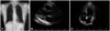
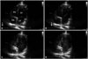
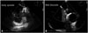
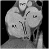
On physical examination, clubbing fingers of both hands were detected (Fig. 1). Chest X-ray finding was normal with cardio-thoracic ratio of 0.4 (Fig. 2A). Electrocardiography revealed normal axis without evidence of cardiac chamber enlargement. Complete blood count showed polycythemia (hemoglobin 19.3 g/dL, hematocrit 56.4%) but JAK2 mutation was negative which suggests polycythemia was secondary change from hypoxemia. Brain magnetic resonance imaging (MRI) showed right cerebellar small embolic nature infarction. No intra- or extra-cranial vascular obstruction was detected. Interestingly, his initial blood gas analysis showed hypoxemia (PaO2 58.1 mm Hg, O2 saturation 90.7% at room air) with mild hypocapnea (PaCO2 27.3 mm Hg) which could not be corrected with oxygenation therapy (PaO2 70.8 mm Hg, O2 saturation 93.6% at 4 L oxygenation by nasal cannula). Putting the laboratory tests together, intra- or extra-cardiac shunt was suspected. However, on initial transthoracic echocardiography (TTE), there were no signs of heart chambers enlargement and no definite intra-cardiac shunt flow was detected (Fig. 2B and C). After the initial work-up, he was on clopidogrel 75 mg daily medication and discharged from neurology department without transesophageal echocardiography (TEE). After 6 months, he readmitted to neurology department with second attack of stroke. The brain MRI revealed acute embolic infarction on right thalamus without large vessel pathology. TTE with bubble test showed rapid filling of LA and left ventricle chambers with agitated bubble simultaneously within 3-4 cardiac cycles after filling right atrium (RA) and right ventricle chambers (Fig. 3, Supplementary movie 1). TEE showed ASD with bidirectional shunt flow (Fig. 4). Left to right shunt was dominant during most of the cardiac cycle, but right to left shunt was also visible during mid to late diastolic period. Heart dynamic computed tomography showed that the opening of IVC was facing toward ASD, guiding direct transmission to the LA via ASD (Fig. 5).
Cardiac catheterization was performed to rule out pulmonary arterial hypertension. Pulmonary to systemic blood flow ratio was 1.3 and mean pulmonary arterial pressure was within normal range (15 mm Hg). Transient trial balloon occlusion of ASD was able to correct hypoxemia without deterioration of hemodynamic collapse (O2 saturation 90% to 98% after balloon occlusion).
The patient preferred surgical repair rather than device closure, and patch closure with bovine pericardium was performed. After the surgery, laboratory finding turned to normal with hemoglobin 12.3 g/dL, hematocrit 40.2%, blood gas analysis of PaO2 86.4 mm Hg, and O2 saturation 96.2% at room air. He was discharged after successful recovery and regularly visiting outpatient clinic without symptom until present.
Discussion
Patients with long standing ASD are in the risk of pulmonary arterial hypertension. Pulmonary hypertension in ASD patient results in elevation of RA pressure and consequently develops predominant right to left shunt. Deoxygenated systemic venous blood flow directly passes through the defect and ejected again during left ventricular systole to systemic arterial circulation.1) These findings make the patient cyanotic and show minimal response to supplemental oxygen.3) Pulmonary hypertension with right to left shunting at rest is the principal contraindication to ASD closure.4) In this case, the patient showed cyanosis with normal pulmonary arterial pressure. Godart et al.2) suggested two theories to explain the development of a right to left shunt with normal pulmonary artery pressure: 1) hemodynamic phenomenon with an interatrial pressure gradient such as RA myxoma, right ventricular infarction, and mechanical ventilation; 2) preferential blood flow streaming from the IVC to the LA through the defect without pressure gradient. Overdeveloped Eustachian valve can guide blood flow from the IVC through the ASD.5) Distortion and tilting of heart structure by aortic aneurysm, pneumonectomy or abdominal surgery is another reason for preferential blood flow from the IVC to the ASD.6)7)8) In the present case, anterolateral deviation of the IVC led direct flow through ASD opening to the LA.
Presence of right to left shunt is an important cause of paradoxical embolism.9) In polycythemia, cerebral blood flow can decrease up to 50% due to increased blood viscosity.10) In this patient, right to left shunt and decreased cerebral flow due to polycythemia might have contributed to recurrent stroke event.
We present an interesting case of ASD with normal pulmonary arterial pressure manifesting with recurrent stroke in young adult. Right to left shunt was due to the unique IVC position directly facing ASD opening. Position of IVC and ASD opening should be considered in cyanotic ASD patients with normal sized heart chambers and pulmonary arterial pressure.

 XML Download