Journal List > J Cardiovasc Ultrasound > v.22(4) > 1018339

Kharwar, Sharma, Sethi, Narain, and Saran: Pulmonary Artery Stenosis due to Lung Carcinoma: A Rare Cause of Dyspnea

Abstract

Acquired bilateral pulmonary artery stenosis in adults due to lung malignancy is infrequently reported. We describe an adult male who presented to us with chief complaints of dyspnea on exertion and one episode of hemoptysis. Two dimensional transthoracic echocardiography with color Doppler showed presence of an extra cardiac mass causing severe extrinsic compression of both the right and left pulmonary artery leading to high pressure severe tricuspid regurgitation and extension of the mass into the left atrium. Three dimensional transthoracic echocardiography clearly delineated the anatomy of the left atrial mass and its surrounding anatomical relationship. The diagnosis of non small cell lung carcinoma was confirmed by multidetector computed tomography (MDCT) and with MDCT guided biopsy with histopathology. Patient succumbed one month later due to an episode of massive hemoptysis.

Introduction

Lung carcinoma is the leading cause of death related to cancer in both men and women through-out the world. Primary lung carcinoma spreading to pulmonary vessels is quite rare and only few individual cases have been reported in the medical literature.1) The prognostic significance of pulmonary vessel invasion by lung carcinoma is unclear and the treatment is challenging. We aim to describe a case of a primary lung carcinoma infiltrating into both the pulmonary arteries and the left atrium who presented to us with dyspnea and hemoptysis.

Case

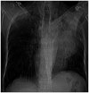
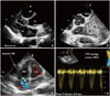
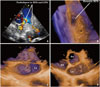
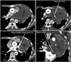
A 50-year-old male presented to us with effort dyspnea of NYHA functional class II for last 3 months. He also had one episode of hemoptysis one month back. General examination was significant for suboptimal nutrition and a prominent jugular venous pulsation. Cardiac auscultation revealed a grade III/VI pansystolic murmur at left 3rd and 4th parasternal region which was increasing in intensity on inspiration. Also there was a grade III/VI ejection systolic murmur in the pulmonary area. Electrocardiogram had evidence of right ventricular pressure overload. Chest X-ray (Fig. 1) showed a large opaque shadow in the upper and middle zone of the left lung which could not be separated from the cardiac silhoute. Also the left hemi-diaphragm was higher than the right hemi-diaphragm. Two dimensional (2D) trans thoracic echocardiography (TTE) showed a large extra-cardiac, echo-dense, non mobile mass near the right ventricular outflow and pulmonary artery region (Fig. 2A and B, Supplementary movie 1 and 2) causing severe extrinsic compression of both the right and the left pulmonary artery resulting into severe high pressure tricuspid regurgitation with a peak velocity of 4.2 m/sec (Fig. 2C and D). There was also an abnormal mass visualized into the left atrium in the parasternal long axis view (Fig. 2A, Supplementary movie 3). Three dimensional (3D) TTE clearly delineated the surface characteristics and the anatomical relationship of the mass in the left atrium (Fig. 3C and D, Supplementary movie 4 and 5) as well as stenosis of the right pulmonary artery caused by the mass (Fig. 3B). Evaluation by multidetector computed tomography (MDCT) showed a large mass (measuring 8.0 × 8.0 × 7.0 cm) in the upper left pulmonary region encroaching upon and causing severe extrinsic compression of both the pulmonary arteries (Fig. 4A and B). The same mass was infiltrating into the left atrium in the form of a pedunculated growth (Fig. 4C and D). Computed tomography guided biopsy revealed non-small cell lung carcinoma (NSCLC) and patient was graded as T4 NSCLC due to in-growth into left atrium. He was started on cisplatin and paclitaxel based chemotherapy, but patient succumbed one month later due to a large bout of hemoptysis.

Discussion

Acquired pulmonary artery stenosis is rarely found after childhood1) and a tumor causing bilateral pulmonary artery compression and involvement of the left atrium is rarely reported. The differential diagnosis for the etiology of the extrinsic pulmonary artery compression includes malignant tumors, infection, cyst, and other benign processes leading to fibrosis in the mediastinum. The treatment is directed towards control of the underlying disease. 2D transthoracic echocardiography plays an important role in evaluation of right heart valvular disease.2) The role of 3D echocardiography for evaluation of intracardiac masses is also evolving.3) In the present case, 2D TTE with color Doppler suggested the diagnosis and 3D TTE was complimentary to 2D TTE. The final diagnosis was ultimately made by MDCT with histopathological confirmation.
In short, malignancy is one of the causes of acquired pulmonary artery stenosis in adults and 2D TTE is an important non invasive tool for detection of hemodynamic compromise resulting from the pulmonary flow obstruction. 3D TTE can be useful in evaluation of cases where tumor has an intracardiac extension.

Figures and Tables

Fig. 1
Chest X-ray PA view showing an opaque shadow in the upper and middle zone of the left lung which is inseparable from the cardiac silhoute. Also note that the left hemi-diaphragm is higher than the right hemi-diaphragm.
jcu-22-209-g001
Fig. 2
Two dimensional transthoracic echocardiography with color Doppler. The parasternal long axis view (A) is showing a mass in the left atrium. The extracardiac mass (indicated by *) is seen to involve the region of the MPA and right and the left pulmonary artery region in the basal short axis view (B). Severe tricuspid regurgitation is seen in the apical four chamber view (C) with a peak TR velocity of 4.2 m/sec on continous wave Doppler (D). Ao: aorta, LA: left atrium, LV: left ventricle, MPA: main pulmonary artery, RV: right ventricle, TR: tricuspid regurgitation, RPA: right pulmonary artery.
jcu-22-209-g002
Fig. 3
Two dimensional transthoracic echocardiography with color Doppler. The basal short axis (A) view with focus on the pulmonary artery showing severe turbulence in RPA and LPA caused by extrinsic compression of a huge mass. Three dimensional transthoracic echocardiography showing stenotic right pulmonary artery caused by the extrinsic compression by the mass (B) and the surface characteristics and anatomical relationship of the intra atrial extension of the mass (indicated by * in C and D). Ao: aorta, LA: left atrium, LPA: left pulmonary artery, RA: right atrium, RPA: right pulmonary artery.
jcu-22-209-g003
Fig. 4
Multidetector computed tomography with contrast showing a large mass (indicated by *) in the upper left pulmonary zone encroaching upon and causing severe extrinsic compression of the RPA and LPA as shown in the axial sections (A and B). The mass has infiltrated into the left atrium in the form of a pedunculated growth as seen in the axial section (C) and in the coronal section (D). AA: ascending aorta, DA: descending aorta, LA: left atrium, LPA: left pulmonary artery, RPA: right pulmonary artery, MPA: main pulmonary artery.
jcu-22-209-g004

References

1. Viseur P, Unger P. Doppler echocardiographic diagnosis and follow-up of acquired pulmonary stenosis due to external cardiac compression. Cardiology. 1995; 86:80–82.
crossref
2. American College of Cardiology. American Heart Association Task Force on Practice Guidelines (Writing Committee to revise the 1998 guidelines for the management of patients with valvular heart disease). Society of Cardiovascular Anesthesiologists. Bonow RO, Carabello BA, Chatterjee K, de Leon AC Jr, Faxon DP, Freed MD, Gaasch WH, Lytle BW, Nishimura RA, O'Gara PT, O'Rourke RA, Otto CM, Shah PM, Shanewise JS, Smith SC Jr, Jacobs AK, Adams CD, Anderson JL, Antman EM, Fuster V, Halperin JL, Hiratzka LF, Hunt SA, Lytle BW, Nishimura R, Page RL, Riegel B. 2006 guidelines for the management of patients with valvular heart disease: a report of the American College of Cardiology/American Heart Association Task Force on Practice Guidelines (writing Committee to Revise the 1998 guidelines for the management of patients with valvular heart disease) developed in collaboration with the Society of Cardiovascular Anesthesiologists endorsed by the Society for Cardiovascular Angiography and Interventions and the Society of Thoracic Surgeons. J Am Coll Cardiol. 2006; 48:e1–e148.
3. Plana JC. Three-dimensional echocardiography to assess intra-cardiac masses. In : Badano LP, Lang RM, Zamorano JL, editors. Textbook of real-time three dimensional echocardiography. London: Springer-Verlag;2011. p. 111–119.

Supplementary movie legends

Movie 1

Two dimensional transthoracic echocardiography. Basal short axis view showing a large mass in the pulmonary artery region with extension into the left atrium. AV: aortic valve, LA: left atrium, PA: pulmonary artery.

Movie 2

Two dimensional transthoracic echocardiography with color Doppler. Basal short axis view showing a large extracardiac mass causing extrinsic compression of the right and the left pulmonary artery as shown by color flow turbulence. AV: aortic valve, RPA: right pulmonary artery, RVOT: right ventricular outflow tract.

Movie 3

Two dimensional transthoracic echocardiography. Parasternal short axis view showing a mass in the left atrium. Ao: aorta, LA: left atrium, LV: left ventricle.

Movie 4

Three dimensional (3D) transthoracic echocardiography. The anatomical details and relationship of the mass in the left atrium can be clearly delineated in this real time 3D view in the basal short axis orientation. AV: aortic valve, LA: left atrium, RA: right atrium.

Movie 5

Three dimensional (3D) transthoracic echocardiography. The anatomical details and relationship of the mass in the left atrium can be clearly delineated in this real time 3D view in the parasternal long axis orientation.
TOOLS
Similar articles