

 PDF
PDF ePub
ePub Citation
 Print
Print


Introduction
Peripartum cardiomyopathy (PPCMP) is a cardiac condition characterized by development of heart failure during the last month of pregnancy or during the first five months of post partum period without any other identifiable cause of heart failure.1) The hypercoagulable state in the pregnancy along with left ventricular (LV) systolic dysfunction predisposes the patient to thromboembolic complications like intraventricular thrombi. Two dimensional (2D) and three dimensional (3D) echocardiography are valuable tools for evaluation of intra-cardiac masses. We report an unusual case of a pedunculated highly mobile LV mass in a patient with PPCMP evaluated by 3D transthoracic echocardiography (TTE) responding to oral anticoagulation.
Case
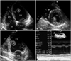
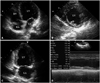
A 30-year-old gravida 4 and para 4 female presented to our department for evaluation of recent onset dyspnea on exertion along with orthopnea. She had delivered a healthy baby 3 weeks ago through normal vaginal delivery. There was no history of pre-existing cardiac illness. On general examination, pulse rate was 110 per minute, blood pressure 120/70 mmHg, respiratory rate of 22 per minute and jugular venous pressure was raised. Cardiac auscultation did not reveal any significant finding and chest examination showed bilateral basilar fine crepitations. The blood investigations including complete blood count, renal function test and liver function tests were within normal range. Electrocardiogram showed sinus tachycardia and chest X-ray showed cardiomegaly along with features of raised pulmonary venous pressure. Evaluation by 2D TTE (Fig. 1, Supplementary movie 1) showed dilatation of all four cardiac chambers and global hypokinesia of the LV. The LV ejection fraction was 32%, LV end diastolic dimension index was 3.05 cm/m2 and M mode LV fractional shortening was 15%. There was a highly mobile pedunculated mass (2.5 × 2.0 cm) attached to the interventricular septum protruding into the LV cavity. There was no history of diabetes, hypertension, preeclampsia, eclampsia, family history of cardiomyopathy or any metabolic disorders that could lead to cardiomyopathy. Hence the diagnosis of PPCMP with intra-cardiac thrombus was considered. 3D TTE evaluation (Fig. 2, Supplementary movie 2) was done to further delineate the anatomy of the LV mass. 3D TTE clearly delineated the attachment of the mass as well as the internal echolucent areas within the mass. Based on the clinical scenario and the 3D TTE finding the mass was suspected to be a thrombus. However, the possibility of cardiac tumour could not be excluded. Because of the mobility of the mass and uncertainty of its nature, the risk of embolization was considered to be high. The patient was advised surgical removal of the mass, but she opted for oral anticoagulation therapy. She was started on standard heart failure therapy along with warfarin (target international normalized ratio 2.0-3.0). Repeat echocardiography 1 month later (Fig. 3, Supplementary movie 3) showed complete dissolution of the LV mass along with an improvement in LV ejection fraction (43%). There was no history of embolization in between. The response to anticoagulation therapy suggested that the pendunculated LV mass was in fact a thrombus.
Discussion
The true incidence of PPCMP is unknown, but it is estimated to be between 1:15000 to 1:1300 deliveries.2) PPCMP has been associated with several risk factors including increased age, gravidity or parity, African origin, toxaemia or hypertension of pregnancy, use of tocolytics, twin pregnancy, obesity and low socioeconomic status.3) The exact etiology of PPCMP is not known and the patient are managed on the lines of standard heart failure regimes. The hypercoagulable state during the pregnancy along with the poor LV systolic function provides a fertile soil for the development of thromboembolic complications including intracardiac thrombi. Up to 53% of cases of PPCMP have been reported to have thromboembolism1)4) although majority have been reported at autopsy or after the clinical occurrence of the embolic episode. Intra cardiac thrombus rarely presents as a pedunculated structure. Cardiac fibroma, cardiac hemangioma, blood cyst and aspergillus mural endocarditits can all present as pedunculated mass in the LV cavity but the LV ejection fraction is usually preserved in these cases. The sensitivity of 2D echocardiography for detecting LV thrombi is 92% to 95% and the specificity is 86% to 88%.5) A retrospective study using surgical confirmation as the gold standard suggested that contrast-enhanced magnetic resonance imaging is superior to TTE and transesophageal echocardiography in both sensitivity and specificity.6) However the time required for the study and its high cost limits its routine clinical use. Real time 3D TTE is a newer technique for evaluation of intra cardiac masses. The 3D data set once acquired, can be cropped and sliced in many different ways and additional information about mass location, shape, attaching interface and relationships with adjacent structures can be derived. In our case, 3D TTE performed better than 2D TTE for exact delineation of 1) the irregular surface characteristics of the mass, 2) the narrow based attachment of the mass via a pedicle to the interventricular septum, and 3) internal echolucency within the mass. The echolucency within the thrombi has been reported to indicate internal clot lysis and liquefaction.7)8) The irregular surface characteristics of the thrombi in our case indicated its recent origin and the internal echolucency predicted its possible response to anticoagulation. A pedunculated LV thrombus that is connected to septum or the LV wall, which is highly mobile and which moves throughout the cardiac cycle, is a risky situation as it has got a high potential for embolization despite adequate anticoagulation.9) Over the years, the primary therapeutic option for such a thrombi has included thrombectomy, anticoagulation and thrombolyis. Still the definitive therapy is controversial and oral anticoagulation has variable success, with resolution rates between 13% and 59%. Very few cases of PPCMP have been reported in the literature where the intra cardiac thrombi have rapidly responded to anticoagulation with complete resolution of the thrombi.10)
Our patient had a pedunculated highly mobile LV mass in the setting of PPCMP. Due to its narrow pedicle and high mobility, the risk of embolization was considered to be very high and patient was advised to undergo surgical removal of the mass. The information obtained from 3D TTE and its response to anticoagulation confirmed the nature of the mass to be a thrombus. Although surgical thrombectomy is the preferred treatment strategy in such cases, our patient responded well to anticoagulation.
 XML Download