Journal List > J Cardiovasc Ultrasound > v.22(2) > 1018363

Kharwar, Dwivedi, Chandra, and Saran: Vanishing Left Ventricular Thrombi in Severe Aortic Stenosis with Dilated Cardiomyopathy
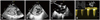
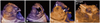
A 20-year-old male presented to our department with complaints of dyspnea on effort for six months. On clinical examination, he had a low volume slow rising pulse, blood pressure of 100/70 mmHg and a raised jugular venous pressure. Cardiovascular examination revealed a downward and outward shifted apical impulse with an ejection systolic murmur of grade IV/VI intensity heard best at the right second intercostal space radiating to both carotids. There were bilateral basilar fine crepitations in chest. Two dimensional transthoracic echocardiography (2D TTE) with color Doppler (Fig. 1, Supplementary movie 1, 2, and 3) showed dilatation of all four cardiac chambers, left ventricle (LV) ejection fraction of 25% and multiple large layered as well as non-layered thrombi in the LV cavity. Aortic valve (AV) was bicuspid with severe aortic stenosis having a peak velocity of 4.2 m/sec. Three dimensional transthoracic echocardiography (3D TTE) clearly showed various details of the multiple thrombi like size, shape, mobility, number, location as well as the internal echolucent areas within the thrombus suggestive of clot lysis (Fig. 2, Supplementary movie 4). Patient denied any form of surgical intervention and was started on oral anticoagulation with warfarin (target INR: 2.0-3.0) along with standard decongestive therapy. A review echocardiography after 8 weeks (Fig. 3) showed complete disappearance of the LV thrombi. LV ejection fraction was almost unchanged.
Congenital bicuspid AV is present in about 1-2% of the population and is more common in males.1) Most bicuspid AV function normally until late in life, although a subset of patients present in childhood or adolescence. Severe aortic stenosis can occur due to the bicuspid AV and if not treated in time can lead to LV dilatation with poor ejection fraction, which in turn can lead to pulmonary hypertension and right ventricular dilatation and dysfunction. In patients with dilated cardiomyopathy, the reported LV thrombus is 10-30%.2) Non-invasive assessment with 2D TTE plays a pivotal role in diagnosis of bicuspid AV with aortic stenosis and LV dysfunction.3) There is a clear advantage of 3D TTE over 2D TTE in the assessment of intra-cardiac masses. Once the images have been acquired, cropping of the data sets can provide a unique view of the interior composition of the mass which can reveal information about its nature. Since the clot lysis begins from inside to outside, 3D TTE reveals this important information by demonstrating areas of echolucency within the clot.4)

Figures and Tables

Fig. 1
Two dimensional transthoracic echocardiography with color Doppler. Multiple large layered as well as non-layered thrombi in LV marked by * are seen in slightly tilted parasternal long axis view (A) and in short axis view at the papillary muscle level (B). Bicuspid AV is clearly visible in parasternal basal short axis view (C) which was the cause of severe aortic stenosis with a peak velocity of 4.2 m/s on continuous wave Doppler (D). AV: aortic valve, LA: left atrium, LV: left ventricle, RA: right atrium, RV: right ventricle.
jcu-22-98-g001
Fig. 2
Real time three dimensional transthoracic echocardiography with volume rendering. The size, shape, number, mobility, surface characteristics, intracardiac location of thrombi can be clearly delineated in various imaging planes (A and B). Echolucency within the thrombi (arrows in C) is seen suggestive of clot lysis beginning from inside to outside. Bicuspid AV can be delineated in three dimensional full volume zoomed mode (D). Thrombi in LV are marked by *. AV: aortic valve, LA: left atrium, LV: left ventricle.
jcu-22-98-g002
Fig. 3
Two dimensional transthoracic echocardiography after 8 weeks of anticoagulation therapy. The multiple thrombi in the LV cavity have completely disappeared as seen in apical four chamber (A), parasternal long (B), and short axis (C) views. The LV ejection fraction is 25% which has relatively unchanged from the previous reading (D). Ao: aorta, LA: left atrium, LV: left ventricle, RA: right atrium, RV: right ventricle.
jcu-22-98-g003

References

1. Nistri S, Basso C, Marzari C, Mormino P, Thiene G. Frequency of bicuspid aortic valve in young male conscripts by echocardiogram. Am J Cardiol. 2005; 96:718–721.
crossref
2. Gottdiener JS, Gay JA, VanVoorhees L, DiBianco R, Fletcher RD. Frequency and embolic potential of left ventricular thrombus in dilated cardiomyopathy: assessment by 2-dimensional echocardiography. Am J Cardiol. 1983; 52:1281–1285.
crossref
3. Visser CA, Kan G, David GK, Lie KI, Durrer D. Two dimensional echocardiography in the diagnosis of left ventricular thrombus. A prospective study of 67 patients with anatomic validation. Chest. 1983; 83:228–232.
crossref
4. Duncan K, Nanda NC, Foster WA, Mehmood F, Patel V, Singh A. Incremental value of live/real time three-dimensional transthoracic echocardiography in the assessment of left ventricular thrombi. Echocardiography. 2006; 23:68–72.
crossref

Supplementary movie legends

Movie 1

Two dimensional transthoracic echocardiography apical 4 chamber view. There is a large thrombus visible inside the left ventricular cavity. Also note the dilatation of all the four cardiac chambers along with poor left ventricular contractility.

Movie 2

Two dimensional transthoracic echocardiography parasternal short axis view. There are multiple layered as well as non-layered left ventricular thrombi along with poor left ventricular contractility.

Movie 3

Three dimensional transthoracic echocardiography modified parasternal long axis view. The left ventricular cavity appears to be full of thrombi. Note the different spin and direction of movement of each thrombus.

Movie 4

Real time three dimensional transthoracic echocardiography with volume rendering. The number, shape, mobility, surface characteristics, intracardiac location of thrombi is clearly delineated. Echolucency, seen within one of the thrombus, is indicative of clot lysis.
TOOLS
Similar articles