

 PDF
PDF ePub
ePub Citation
 Print
Print


Introduction
Aortic stenosis (AS) is a highly and increasingly prevalent condition that has become a major health concern.1) Characteristically, patients present a long latent asymptomatic period where the risk of sudden death is low, even with severe AS: i.e., peak aortic velocity ≥ 4 m/s, mean aortic gradient ≥ 40 mmHg and aortic valve area ≤ 1.0 cm2 or ≤ 0.60 cm2/m2. However, the risk of sudden death increases dramatically when symptoms appear justifying the European Society of Cardiology/European Association of Cardiothoracic Surgery (ESC/EACTS) to underline the role of exercise testing to clarify symptomatic status in patients with severe AS.2) On the other side, "truly" asymptomatic patients undergoing early aortic valve replacement (AVR) may have better outcome compared to medically treated patients.3) Hence, the determination of individual risk factors of rapid clinical deterioration could help identify patients who may benefit most from early elective surgery. Accordingly, recent studies have demonstrated that exercise echocardiography can provide incremental prognostic value over resting echocardiography and exercise testing.4)5)6) Exercise echocardiography is useful because it allows assessing clinical, hemodynamic and functional adaptation responses during exercise, which are directly linked to functional status, degree of ventriculo-arterial coupling and left ventricular (LV) myocardial reserve. The purpose of this article is to describe the role of exercise testing and echocardiography in the management of asymptomatic patients with severe AS and preserved LV ejection fraction.
Exercise Testing
Protocol
A complete clinical evaluation to rule out the presence of symptoms and to identify potential contraindications is essential before submitting patients to exercise testing. Contraindications to exercise testing include: clear indications for AVR (i.e., symptomatic severe AS), uncontrolled hypertension (systolic pressure > 220 mmHg or diastolic pressure > 110 mmHg), uncontrolled or symptomatic arrhythmias, physical or mental disability with the inability to adequately exercise and systemic disease limiting exercise performance.7) A symptom-limited exercise test performed with the goal to reach at least 85% of the age-predicted heart rate is recommended. Treadmill or semi-supine bicycle exercise testing can be used. Safety of both techniques has already been demonstrated and complications remain low under appropriate supervision and monitoring.8)9) Treadmill exercise is more commonly used in North America and is realized according to the ACC/AHA practice guidelines using a modified Bruce protocol.10) In contrast, semi-supine ergometer with a tilting table is the preferred approach in Europe reducing the potential risk of hemodynamic collapse compared to treadmill test.11) Patients should continue their usual medications, as abnormal results on suboptimal therapy may be confusing for management decisions. The workload should be adjusted for each patient, i.e., beginning at 50 W with an increase of 25 W every 2 minutes for a young patient versus starting at 25 W with an increase of 10 W every 2 minutes for an older patient. Appearance of symptoms should be assessed regularly, and blood pressure, heart rate and 12-lead electrocardiography should be monitored continuously during the examination. Exercise testing should be interrupted when the target heart rate is reached or if the patient presents typical chest pain, limiting breathlessness, dizziness, hypotension (drop in systolic blood pressure ≥ 20 mmHg), significant ventricular arrhythmia or muscular exhaustion.
Interpretation
Symptomatic status can be difficult to establish because patients may minimize or deny their symptoms or reduce their level of physical activity to avoid them, especially in elderly. Then, exercise testing can be useful to unmask symptoms in patients with severe AS. Approximately one third of patients who claim to be asymptomatic will develop symptoms on exercise testing.12)13) However, the occurrence of rapidly reversible dyspnea at high workloads should not be interpreted as abnormal. Interpretation of exercise testing could be limited in elderly population. In fact, positive predictive value in patients > 70 years old has been shown to be significantly lower than in younger patients (56% compared to 79%) with similar negative predictive values.13) According to this, a negative exercise test should be reassuring, but a positive exercise test may lack of specificity due to frequent comorbidities in elderly patients. Also, ST segment depression may not improve the positive predictive value of exercise testing, particularly in patients with concomitant coronary artery disease.13) Generally, exercise testing is considered positive when the patient presents ≥ 1 of the following criteria: angina, limiting dyspnea at low workloads, syncope or near-syncope, ≥ 2 mm horizontal or down-slopping ST segment depression, drop or ≥ 20 mmHg rise in systolic blood pressure or complex ventricular arrhythmias.7)
Impact on clinical decision-making
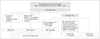
Symptom-limited exercise testing can add important prognostic value.14) For this reason, exercise testing is strongly advocated by the ESC/EACTS (Class I recommendation) in asymptomatic patients with severe AS.2) In fact, a positive exercise test has been shown to predict the rapid onset of symptoms, the occurrence of cardiac death and the need for AVR. To note, exercise-induced dizziness have the highest positive predictive value for the occurrence of symptoms during follow-up.12) In a meta-analysis examining 491 patients, a negative exercise test was associated with no sudden death while 5% of patients with a positive result presented with sudden death during 12-month follow-up. Also, 21% of patients with a negative exercise test had adverse cardiac events compared to 66% of patients with a positive result.14) Therefore, current ESC/EACTS guidelines recommend AVR in asymptomatic patients with severe AS who develop symptoms during exercise testing (ESC/EACTS, Class I) or a fall in systolic blood pressure below baseline value (ESC/EACTS, Class IIa) (Fig. 1).2)
Exercise Echocardiography
Protocol
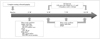
In valvular heart disease, an experienced sonographer or cardiologist should perform exercise echocardiography. Both types of exercise test can be used. Treadmill allows only post-exercise imaging limiting the accuracy of measurements compared to semi-supine cyclo-ergometer permitting optimal image acquisitions during each step of exercise testing. Comprehensive resting echocardiography should be performed in the same position as during the exercise testing. Echocardiographic parameters related to the severity of AS, the consequences on the LV and the systolic pulmonary arterial pressure (SPAP) should be recorded throughout the test. To note, the ratio of early diastolic mitral inflow velocity to early diastolic annulus velocity (E/e' ratio) should be measured before E and A wave fusion appearing at higher heart rates (usually > 100-110 bpm) (Fig. 2).
Interpretation
Changes in mean aortic gradient
Regardless of resting AS severity, an increase in mean aortic pressure gradient by ≥ 18-20 mmHg during exercise, which occur in about one-third of patients, is associated with an increased risk of cardiac-related events.4)6) In fact, an exercise-induced increase in mean aortic pressure gradient by ≥ 20 mmHg has been identified as the most powerful predictor of poor outcomes, even after adjustment for age, exercise LV ejection fraction and resting mean aortic gradient.6) Such an increase in mean aortic pressure gradient partly reflects the presence of a rigid and non-compliant aortic valve.15) Moreover, the kinetics of change in mean aortic pressure gradient needs to be assessed as a rapid increase may indicate the presence of a more severe disease.
LV systolic functional reserve
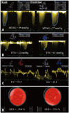
Assessment of LV function during exercise can also provide incremental prognostic information. Limited contractile reserve (i.e., a decrease or a limited increase in LV ejection fraction) has been shown to be associated with an abnormal hemodynamic response to exercise, the development of symptoms and cardiovascular death.16)17) Limited contractile reserve may represent a more advanced disease process with subclinical intrinsic myocardial damage. The afterload mismatch and the exhausted coronary flow reserve with consequent subendocardial ischemia and myocardial fibrosis could contribute to limited functional LV adaptation during test.18) As previously reported, standard LV ejection fraction measurements are relatively insensitive to detect early forms of myocardial dysfunction and assessment of LV longitudinal function seems to be more powerful in predicting the occurrence of symptoms, exercise intolerance and outcome in AS.19)20) Longitudinal function can be studied using tissue Doppler imaging with pulsed-wave Doppler at the mitral annulus measuring s' velocities or using 2-dimentional speckle tracking analyzing global longitudinal strain (GLS) (Fig. 3). In a cohort of AS patients, a smaller increase in s' velocities after exercise was associated with a lower exercise capacity and a lower exercise-induced increase in systolic blood pressure.21) According to strain analysis, Lafitte et al.19) demonstrated that patients with an abnormal exercise response had a significant lower resting GLS and basal longitudinal strain (BLS) compared to patients with a normal exercise test (respectively -14.7% vs. -19.3% and -10.7% vs. -14.4%). Cutoff values of -18% for GLS and -13% for BLS were able to predict an inadequate exercise response with a sensitivity/specificity of 68/75% (GLS) and 77/83% (BLS). Moreover, Donal et al.22) reported that exercise-induced changes in LV GLS were lower in AS patients with an abnormal exercise response. In multivariable analysis, a lower resting GLS, a higher exercise-induced increase in mean aortic pressure gradient and a smaller exercise-induced change in GLS were predictors of a positive exercise test.
LV diastolic functional reserve
In patients with severe AS, the chronically increased afterload is compensated by LV hypertrophy. At a later stage, this adaptive response may be no longer sufficient to mitigate the clinical impact of an increase in LV filling pressure. The E/e' ratio is recognized as a noninvasive estimate of LV filling pressure. At rest, a cutoff value of E/e' > 13 has been shown to identify LV end-diastolic pressure > 15 mmHg with a sensitivity of 93% and a specificity of 88%.23) At exercise, cutoff values of E/e' between 13 and 15 were correlated with elevated LV end-diastolic pressure during exercise and reduced exercise capacity in patients without valvular disease.24)25) However, these exercise values have not been validated in AS patients.
Changes in systolic pulmonary artery pressure
In asymptomatic patients, exercise echocardiography may unmask dynamic pulmonary hypertension (PHT). In fact, an early exercise-induced increase in SPAP is in favour of a lack of pulmonary vascular adaptation with a high resistance and a low compliance reflecting a more severe disease. In a recent prospective study, exercise-induced PHT, defined as SPAP > 60 mmHg, was associated with a 2-fold increase risk of cardiac events at 3-year follow-up in asymptomatic patients with severe AS and preserved LV ejection fraction. Also, in multivariable model, exercise PHT was identified as an independent predictor of cardiac events, even when exercise-induced changes in mean aortic gradient were added.5)
Impact on clinical decision-making
Because of its incremental prognostic value, exercise echocardiography may be useful to improve risk stratification and identify patients who may benefit from an early surgery. According to this, the ESC/EACTS recently added a recommendation based on exercise echocardiography. In asymptomatic patients with severe AS and preserved LV ejection fraction, AVR may be considered in patients with an exercise-induced increase in mean aortic pressure gradient > 20 mmHg (Class IIb).2) Otherwise, patients with a significant increase in mean aortic pressure gradient, a limited LV contractile reserve or PHT during exercise could be referred to a dedicated Heart Valve Clinic for a closer follow-up (3-6 months) including reevaluation of symptoms and repeated exercise echocardiography.26) Conversely, patients without markers of poor prognosis may be safely reassessed every 12 months (Fig. 1). However, prospective clinical trials are needed to support the widespread use of exercise echocardiography in the routine management of asymptomatic AS.
Conclusion
In AS, exercise echocardiography can help unmask falsely asymptomatic patients as well as patients with abnormal hemodynamic and functional adaptation to exercise who may benefit from early elective AVR. Large-scale studies are needed to examine the impact of exercise echocardiography results on post-operative outcome.
 XML Download