Journal List > J Cardiovasc Ultrasound > v.21(4) > 1018297

Moustafa, Al Shanawani, Patton, Alvarez, Zuhairy, AlMoukirish, and Mookadam: Early Complication of Mustard Procedure after Late Repair
A 25-year-old woman with complete transposition of the great arteries presented for routine follow-up one year post Mustard procedure. She previously underwent balloon atrial septostomy (Rashkind procedure) early after birth, but her family declined further surgery. She developed progressive pulmonary hypertension early in life, shortness of breath on moderate exertion, followed by progressive exercise intolerance and desaturation (80-85%) over the preceding two years. Pre-operative pulmonary pressure was estimated at approximately 90 mmHg by cardiac catheterization. Subsequently, she underwent a palliative Mustard procedure. Recovery was uneventful with improvement of her functional status and oxygen saturation between 90-95%.
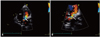
Routine transthoracic echocardiogram (TTE) one year post-procedure revealed a severely dilated, hypertrabeculated systemic right ventricle with mild systolic dysfunction and normal sub-pulmonary left ventricular size and systolic function. Additionally, color Doppler showed turbulence at the junction of the upper and lower limbs of the systemic venous baffles suggestive of significant stenosis without baffle leak (Fig. 1, Supplementary movie 1-3). Cardiovascular magnetic resonance confirmed the TTE findings of significant narrowing of the superior vena cava (SVC) and inferior vena cava (IVC) baffles at the venoatrial junction. The pulmonary venous portion of the baffle was patent. The main pulmonary artery and both branches were severely dilated (Fig. 2, Supplementary movie 4-7).
Due to the presence of significant narrowing of the SVC/IVC baffles, cardiac catheterization was recommended to assess the gradients across the stenotic areas and to potentially perform balloon angioplasty/stenting. However, the patient declined as she felt noticeable improvement of her symptoms following the Mustard procedure.
Herein we present a rare case describing a known complication of Mustard procedures as early as one year post-operatively, in addition to the Mustard procedure itself, which was delayed until 24 years of age. The etiology of baffle stenosis post-Mustard procedure is not clear however, speculations include difficult sizing of the baffles in adult heart, localized scarring of the patch at the suture line or perhaps anatomical distortion. Surprisingly, the patient had an improvement in symptoms after the procedure despite this significant subsequent obstruction of the systemic venous pathways.1-3)

Figures and Tables

Fig. 1
Transthoracic echocardiogram apical 4-chamber view (A) with zoom mode (B) demonstrating the connections of the systemic venous circulation with significant color turbulence suggestive of baffle stenosis at the veno-atrial junction (arrow). LV: left ventricle, PVA: pulmonary venous atrium, RV: right ventricle.
jcu-21-200-g001
Fig. 2
A: Cardiovascular magnetic resonance 4-chamber steady-state free precession (SSFP) image showing the trabeculated dilated systemic RV and patent pulmonary venous baffle (arrow). B: Cardiovascular magnetic resonance coronal SSFP image showing the typical discordant ventriculararterial relationship in complete transposition of the great arteries with the AO arising from the RV and MPA arising from the LV. C: Cardiovascular magnetic resonance axial SSFP image demonstrating severely dilated main PA and both branches. D: Cardiovascular magnetic resonance coronal SSFP image revealing significant stenosis of both superior vena cava (long arrow) and inferior vena cava (short arrow) limbs of the Mustard baffle at the veno-atrial junction. LV: left ventricle, PVA: pulmonary venous atrium, RV: right ventricle, AO: aorta, PA: pulmonary artery, LPA: left pulmonary artery, MPA: main pulmonary artery, RPA: right pulmonary artery.
jcu-21-200-g002

References

1. Chatelain P, Meier B, Friedli B. Stenting of superior vena cava and inferior vena cava for symptomatic narrowing after repeated atrial surgery for D-transposition of the great vessels. Br Heart J. 1991; 66:466–468.
crossref
2. Cohen MD, Johnson T, Ramrakhiani S. MRI of surgical repair of transposition of the great vessels. AJR Am J Roentgenol. 2010; 194:250–260.
crossref
3. Kouchoukos NT, Blackstone EH, Doty DB, Hanley FL, Karp RB. Kirklin/Barratt-Boyes cardiac surgery. 3rd ed. Philadelphia: Churchill Livingstone;2003. p. 1439–1507.

Supplementary movie legends

Movie 1

Transthoracic echocardiogram apical 4-chamber view showing dilated systemic ventricle with mild systolic dysfunction and normal sub-pulmonary ventricular size and systolic function.

Movie 2

Transthoracic echocardiogram apical 4-chamber view (2) with zoom mode (3) demonstrating the connections of the systemic venous circulation with significant color turbulence suggestive of baffle stenosis at the veno-atrial junction.

Movie 3

Transthoracic echocardiogram apical 4-chamber view (2) with zoom mode (3) demonstrating the connections of the systemic venous circulation with significant color turbulence suggestive of baffle stenosis at the veno-atrial junction.

Movie 4

Cardiovascular magnetic resonance coronal steady-state free precession cine image showing normal sub-pulmonary left ventricle size and systolic function with the dilated main pulmonary artery arising from it.

Movie 5

Cardiovascular magnetic resonance coronal stack steady-state free precession cine image revealing the heavily trabeculated, dilated systemic ventricle (with mild systolic dysfunction), the aorta arising from the right ventricle and main pulmonary artery arising from the left ventricle, and significant stenosis of both superior vena cava/inferior vena cava limbs of the Mustard baffle at the veno-atrial junction.

Movie 6

Cardiovascular magnetic resonance sagittal (6) and short axis (7) stack steady-state free precession cine image demonstrating both ventricles side by side with the typical discordant ventricular-arterial relationship.

Movie 7

Cardiovascular magnetic resonance sagittal (6) and short axis (7) stack steady-state free precession cine image demonstrating both ventricles side by side with the typical discordant ventricular-arterial relationship.
TOOLS
Similar articles