

 PDF
PDF ePub
ePub Citation
 Print
Print


Introduction
Double atrial septum is extremely rare congenital anomaly, which has double-walled atrial septum that distinguishes midline interatrial chamber between the two atria.1)2) This interatrial space sometimes distinguished from left atrium by septum primum, otherwise from right atrium by accessory septal structure.2)3) In some aspects, the accessory septal structure distinguishes this space from right atrium, it is presumed it might be persistent left ventricular (LV) valve attached to sinus venosus in fetal period.4) And clinically, most cases of this anomaly are asymptomatic unless manifest as thromboembolic complications,1)5) such as stroke, or transient ischemic attack, that thrombus may be originated from this interatrial space. To date, there has been no case report in Korea, we encountered and reported a rare case of incidentally found isolated double atrial septum by transthoracic echocardiography (TTE).
Case
A 69-year-old male was consulted to cardiology from otolaryngology for pre-operative cardiac evaluation. He had a history of hypertension with no event of cerebrovascular accidents. And he had an operation schedule for oral cavity cancer involving right buccal mucosa (T4N0M0, in TNM staging). His general condition looked poor because of oral cavity malignancy and concurrent chemotherapy. His blood pressure was 103/62 mmHg and pulse rate was 95 bpm with regular heart beat. Chest X-ray showed mild pulmonary edema and bilateral pleural effusion 1 week ago, but improving with negative volume control.
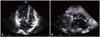
Two-dimensional echocardiography showed normal LV systolic function with LV ejection fraction about 56%, concentric hypertrophy of LV and slightly enlarged left atrial (LA) chamber with LA volume index 55 mL/m2. Apical 4 chamber (Fig. 1A) and parasternal views showed fibrothickened with calcification of mitral valve and aortic valve due to rheumatic heart disease with moderate mitral stenosis (mitral valve area 1.55 cm2, mean diastolic pressure gradient 5.5 mmHg), moderate aortic stenosis (aortic valve area 1.21 cm2, peak/mean systolic pressure gradient 34/19 mmHg) and interatrial septal aneurysm. It also showed mild pulmonary hypertension with 41 mmHg of right ventricular systolic pressure, and inferior vena cava plethora suggestive diastolic heart failure.
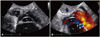
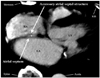
On standard parasternal and apical views, there was no definite abnormal finding at interatrial septum except aneurismal change of interatrial septum (Fig. 1A). However, on subcostal view, there was 3.2 × 1.0 cm sized, crescent shaped, echo-free space was observed between two atria (Fig. 1B). This echo-free space was located in center of interatrial septum and consisted of parallel atrial septal structure (Fig. 2A). It was highly suspicious of double atrial septum with persistent interatrial space. There is no evidence of thrombus in this space and color Doppler echocardiography revealed communicating flow between this space and LA and small amount of left-to-right shunt flow suggesting patent foramen ovale (PFO) (Fig. 2B). The computed tomography (CT) also identified double-layered parallel atrial septal structure with persistent interatrial space, showing contrast enhancement (Fig. 3). We recommended further evaluation for double atrial septum including transesophageal echocardiography (TEE) but patient refused because of oral cavity lesion and had surgery (wide excision of buccal mucosa cancer) at otolaryngology. He was prescribed aspirin and has followed up uneventfully to date.
Discussion
Double atrial septum is extremely rare atrial septal anomaly forms interatrial space distinguished between two atria by parallel double-layered atrial septal structure. This interatrial space usually communicates with left atrium via PFO, and with right atrium via accessory atrial septal fenestration (ASF).2) And these two passages (PFO and ASF) are usually formed in different level, such as superior and inferior.2)
In fetal period, PFO permits right-to-left shunt flow, but double-layered atrial septal structure can disturb this shunt flow therefore, underdevelopment of LV can be frequently combined.2) It can be also affected with LV, LA, mitral valve and pulmonary venous structures in patients with left heart hypoplasia, but these findings were not observed in this case. Roberson et al.2) reported 4 cases of double atrial septum, among these, 2 cases were accompanied with LV hypoplasia, but accurate incidence of LV hypoplasia in patients with double atrial septum was not revealed yet.
Fig. 2A shows accessory atrial septal tissue attached to septum primum and this flap is the finding distinguished from elongated atrial septum secundum. But unfortunately, transesophageal echocardiography was not done in this patient, so it is not obvious whether this double layered atrial septal structure is not elongated atrial septum secundum indeed.6)
Our patient showed moderate mitral stenosis and aortic stenosis, but their etiology was thought to be rheumatic heart disease. And there is no evidence of left heart hypoplasia, including pulmonary venous obstruction. Although we could not perform TEE because of his poor general condition and oral cavity malignancy, but we identified this double-layered parallel atrial septal structure with persistent interatrial space by CT scan. We could not find double atrial septum in standard TTE view however, we could clearly demonstrate double atrial septum with interatrial space in subcostal view. Therefore, routine evaluation of subcostal view of TTE can be useful for the detection of congenital atrial septal malformation.
In conclusion, we described firstly in Korea, an extremely rare case of isolated double atrial septum with persistent interatrial space diagnosed by 2 dimensional TTE. We suggest that careful evaluation of interatrial septum at subcostal view is essential for finding rare interatrial septal anomaly.
 XML Download