Journal List > J Cardiovasc Ultrasound > v.21(3) > 1018284

Kim, Yang, Kim, Kim, Choi, Bae, Lee, Park, Cho, and Chae: Recurrent Syncope Episodes and Exercise Intolerance in Hypertrophic Cardiomyopathy Combined with Atrioventricular Conduction Disturbance

Abstract

A 30-year-old female patient with known hypertrophic cardiomyopathy (HCMP) was admitted for recurrent syncope episodes. Electrocardiogram (ECG) showed 2 : 1 atrioventricular (AV) block. Stress echocardiography with bicycle showed high grade AV block at high stage of the exercise associated with exercise intolerance and dyspnea. Twenty-four hour ECG monitoring also revealed high grade AV block and 1 episode of non-sustained ventricular tachycardia. Implantable cardioverter/defibrillator-pacemaker (ICD-P) was inserted. After implantation of ICD-P, conduction disturbance and exercise intolerance were improved. AV block is a rare complication HCMP. There are just a few case reports that present symptoms caused by conduction disturbance in HCMP. This case describes repeated syncope episodes and exercise intolerance caused by conduction disturbance during exercise in HCMP patient. For evaluating the cause of syncope in HCMP, stress echocardiography can be helpful to understand the probable mechanism of syncope.

Introduction

Syncope or presyncope occurs approximately 25% of patients with hypertrophic cardiomyopathy (HCMP). Recurrent syncope is known as one of the risk factors of sudden cardiac death.1) To give proper management and prevent sudden cardiac death, it is important to know the mechanism causing loss of consciousness in patients with HCMP. Among various etiologies, bradyarrhythmia such as atrioventricular (AV) conduction disturbance, a relatively rare complication associated with HCMP, also can make patient suffer from syncope or presyncope.

Case

A 30-year-old female was referred to the cardiology center due to recurrent syncope episodes and aggravated shortness of breath for a month. She was diagnosed as HCMP 7 years ago, and had been taken atenolol irregularly since then. There was no previous medical history and no family history of any cardiac disease. The first syncope occurred in 2007, during discontinuation of medication. After experienced recurrent syncope episodes, she visited another medical center for evaluation of loss of consciousness. Cardiac evaluation, including conventional echocardiography and 24-hour ambulatory electrocardiogram (ECG) monitoring, couldn't reveal the cause of repeated syncope. The neurologic exams for differential diagnosis showed no evidence of seizure disorder or any other diseases, inducing loss of consciousness. She restarted atenolol for HCMP. However, despite medication, symptoms developed more frequently, combined with shortness of breath and exercise intolerance. In 2012, she visited our cardiology clinic for further evaluation and management of worsening symptoms under medical treatment.
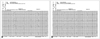
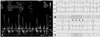
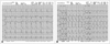
Initial ECG showed 2 : 1 AV block with 36 beats/min of the ventricular rate (Fig. 1A). The baseline two-dimensional echocardiography showed HCMP with asymmetric septal hypertrophy (septal wall thickness during diastole 16 mm) and systolic anterior motion of mitral valve (Fig. 2). After discontinuation of the previous medication (atenolol), follow-up ECG showed normal sinus rhythm (Fig. 1B). To evaluate the cause of recurrent syncope, additional studies including stress echocardiography, 24-hour ambulatory ECG and treadmill test were performed. During the stress echocardiography with bicycle exercise, when reached 50 watts of workload stage, mitral inflow pattern showed multiple spikes during a late filling phase by atrial contraction accompanied with high grade AV block on ECG monitoring (Fig. 3A). Patient suffered from exhaustion and shortness of breath. The baseline blood pressure was 91/59 mmHg (systole/diastole blood pressure) and 46 beats/min of the heart rate, but there was no hypotensive response representing dynamic left ventricular outflow tract obstruction. Simultaneous 24-hour ECG also showed conduction disturbance (including 3 : 1 and 4 : 1 AV block) during the exercise and additionally disclosed 1 episode of non-sustained ventricular tachycardia (NSVT) (Fig. 3B and C). Subsequent treadmill test also revealed high grade AV block with dyspnea and exhaustion at 7.0 METS of exercise under Bruce protocol (Fig. 4A).
Although there was no syncope event when ECG monitoring records high grade AV block or NSVT in both 24-hour ECG and stress test, we thought this conduction disturbance and arrhythmia could play a role in repeated loss of consciousness and exercise intolerance. According to the guideline for risk stratification of sudden cardiac death in HCMP, based on her unexplained syncope episodes, it is reasonable to treat this patient with implantable cardioverter/defibrillator (ICD).2) Patient underwent elective ICD with dual-chamber pacemaker implantation. After that, patient showed no more conduction disturbance at high stage of workload and improved exercise capacity of 12.8 METS under Bruce protocol without any distress in repeated stress test (Fig. 4B).

Discussion

Among the patients with HCMP, about 25-30% of patients experience symptoms of fainting, dizziness or impaired consciousness.2)3) A history of recurrent syncope episodes is one of the predictable risk factors for sudden cardiac death in younger patients.4-6)
Syncope in HCMP can be explained by two underlying mechanisms: hemodynamic mechanism and arrhythmic complications. Among the arrhythmic causes for syncope, paroxysmal atrial fibrillation is the most common cause that induces loss of consciousness.7)8) Unlike premature ventricular complex or NSVT, which is usually not associated with syncope,9) sustained ventricular tachycardia can be the cause of syncope and even sudden cardiac death in HCMP.10) However, besides these tachyarrhymia, bradyarrhythmia such as AV conduction disturbance also cause syncope or pre-syncope in HCMP.11) There are only a very few case reports of HCMP combined with AV block causing syncope both in pediatric and adult patients.12-16)
Despite repeated evaluation, including 24-hour ECG monitoring, conventional two-dimensional echocardiography, and invasive electrophysiological study, probable mechanism causing syncope can be identified only in a limited number of cases.11) However, it is important to understand the cause of syncope in HCMP to avoid risk of sudden cardiac death and to give proper management for each different condition. Medications, such as beta-blocker or calcium channel blocker, usual management for HCMP according to the guideline,2) might aggravate symptoms of patients with conduction disturbance. Stress echocardiography can be helpful to evaluate the cause of syncope in HCMP patients, which provoke exercise-induced hemodynamic change or any arrhythmic complication, either tachyarrhythmia or bradyarrhythmia causing various symptoms.11)
This case describes repeated syncope episodes and exercise intolerance caused by conduction disturbance during exercise in HCMP patients. Rarely complicated in HCMP, physicians should keep in mind the probability of conduction disorder as a cause of syncope.

Figures and Tables

Fig. 1
Baseline electrocardiogram (ECG) showed 2 : 1 atrioventricular conduction block with 36 beats/min of ventricular rate (A) and follow-up ECG after discontinuation previous medications showed normal sinus rhythm (B).
jcu-21-148-g001
Fig. 2
Baseline echocardiogram showed asymmetric septal hypertrophy of left ventricle in parasternal view (A), systolic anterior motion of mitral valve presented by M-mode (B), and left ventricular outflow tract (LVOT) flow pattern with dagger shape and resting pressure gradient via LVOT (C).
jcu-21-148-g002
Fig. 3
Simultaneous stress echocardiogram with abnormal mitral inflow pattern with multiple peaks in atrial phase (A), high grade atrioventricular block (B) and non-sustained ventricular tachycardia in 24-hour electrocardiogram monitoring (C) during exercise.
jcu-21-148-g003
Fig. 4
Initial treadmill test revealed high grade atrioventricular block (A). After implantable cardioverter/defibrillator-pacemaker with ventricular pacing follow-up treadmill test (B), electrocardiogram showed no more conduction disturbance or ventricular arrhythmia during the exercise.
jcu-21-148-g004

References

1. Kofflard MJ, Ten Cate FJ, van der Lee C, van Domburg RT. Hypertrophic cardiomyopathy in a large community-based population: clinical outcome and identification of risk factors for sudden cardiac death and clinical deterioration. J Am Coll Cardiol. 2003; 41:987–993.
crossref
2. American College of Cardiology Foundation/American Heart Association Task Force on Practice; American Association for Thoracic Surgery; American Society of Echocardiography; American Society of Nuclear Cardiology; Heart Failure Society of America; Heart Rhythm Society; Society for Cardiovascular Angiography and Interventions; Society of Thoracic Surgeons, Gersh BJ, Maron BJ, Bonow RO, Dearani JA, Fifer MA, Link MS, Naidu SS, Nishimura RA, Ommen SR, Rakowski H, Seidman CE, Towbin JA, Udelson JE, Yancy CW. 2011 ACCF/AHA guideline for the diagnosis and treatment of hypertrophic cardiomyopathy: a report of the American College of Cardiology Foundation/American Heart Association Task Force on Practice Guidelines. J Thorac Cardiovasc Surg. 2011; 142:e153–e203.
3. Elliott PM, Poloniecki J, Dickie S, Sharma S, Monserrat L, Varnava A, Mahon NG, McKenna WJ. Sudden death in hypertrophic cardiomyopathy: identification of high risk patients. J Am Coll Cardiol. 2000; 36:2212–2218.
crossref
4. Maron BJ, Bonow RO, Cannon RO 3rd, Leon MB, Epstein SE. Hypertrophic cardiomyopathy. Interrelations of clinical manifestations, pathophysiology, and therapy (2). N Engl J Med. 1987; 316:844–852.
5. McKenna W, Deanfield J, Faruqui A, England D, Oakley C, Goodwin J. Prognosis in hypertrophic cardiomyopathy: role of age and clinical, electrocardiographic and hemodynamic features. Am J Cardiol. 1981; 47:532–538.
crossref
6. McKenna WJ, Franklin RC, Nihoyannopoulos P, Robinson KC, Deanfield JE. Arrhythmia and prognosis in infants, children and adolescents with hypertrophic cardiomyopathy. J Am Coll Cardiol. 1988; 11:147–153.
crossref
7. Olivotto I, Cecchi F, Casey SA, Dolara A, Traverse JH, Maron BJ. Impact of atrial fibrillation on the clinical course of hypertrophic cardiomyopathy. Circulation. 2001; 104:2517–2524.
crossref
8. Robinson K, Frenneaux MP, Stockins B, Karatasakis G, Poloniecki JD, McKenna WJ. Atrial fibrillation in hypertrophic cardiomyopathy: a longitudinal study. J Am Coll Cardiol. 1990; 15:1279–1285.
9. Nienaber CA, Hiller S, Spielmann RP, Geiger M, Kuck KH. Syncope in hypertrophic cardiomyopathy: multivariate analysis of prognostic determinants. J Am Coll Cardiol. 1990; 15:948–955.
crossref
10. Spirito P, Bellone P, Harris KM, Bernabo P, Bruzzi P, Maron BJ. Magnitude of left ventricular hypertrophy and risk of sudden death in hypertrophic cardiomyopathy. N Engl J Med. 2000; 342:1778–1785.
crossref
11. Williams L, Frenneaux M. Syncope in hypertrophic cardiomyopathy: mechanisms and consequences for treatment. Europace. 2007; 9:817–822.
crossref
12. Rosen KL, Cameron RW, Bigham PJ, Neish SR. Hypertrophic cardiomyopathy presenting with 3rd-degree atrioventricular block. Tex Heart Inst J. 1997; 24:372–375.
13. Przybojewski JZ, van der, Ellis GC, Tiedt FA. Hypertrophic cardiomyopathy complicated by complete heart block. Case report and review of the literature. S Afr Med J. 1984; 66:847–855.
14. Thongtang V, Panchavinin P, Chaithiraphan S. Familial hypertrophic cardiomyopathy associated with spontaneous complete heart block. J Med Assoc Thai. 1991; 74:301–305.
15. Tamura M, Harada K, Ito T, Enoki M, Takada G. Abrupt aggravation of atrioventricular block and syncope in hypertrophic cardiomyopathy. Arch Dis Child. 1995; 73:536–537.
crossref
16. Desai DM, Bhat GS, Daxini BV, Sharma S. Complete heart block as a cause of syncope in hypertrophic cardiomyopathy. J Assoc Physicians India. 1991; 39:965–966.
TOOLS
Similar articles