

 PDF
PDF ePub
ePub Citation
 Print
Print


Introduction
Subaortic membrane is an uncommon cause of the left ventricular outflow tract (LVOT) obstruction. It is important to distinguish a dynamic LVOT obstruction from fixed LVOT obstruction by a subaortic membrane. Transthoracic echocardiography (TTE) could miss the subaortic membrane close to the aortic valve; transesophageal echocardiography (TEE) could finely visualize subvalvular and supravalvular structures and help to find the other cause of LVOT obstruction such as subaortic membrane.
We report a case of patient who had a flail subaortic membrane with dynamic LVOT obstruction misdiagnosed as obstructive hypertrophic cardiomyopathy (HCMP) with dynamic LVOT obstruction; the subaortic membrane was not seen initially on TTE, but identified by TEE and cardiac catheterization.
Case
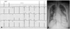
A 67-year-old female presented to our hospital with a symptom of gradually aggravated dyspnea. Clinical examination confirmed the grade 3/6, subaortic, midsystolic murmur and increased respiration rate of 28 breaths/min. A 12-lead electrocardiography showed left ventricular hypertrophy in voltage criteria. A chest radiograph demonstrated marked cardiomegaly with pulmonary edema (Fig. 1).
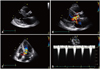
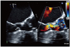
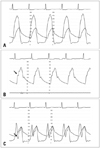
Eight years ago, the patient had come to our hospital with similar symptoms. On TTE, the LV interventricular septal wall thickness and LV posterior wall thickness were 15 mm and 10 mm at diastolic phase, respectively, and papillary muscle was hypertrophied. There was no significant calcification, thickening or motion limitation of aortic valve to increase flow velocity. Continuous wave (CW) Doppler spectrum did not show late peaking appearance but symmetrical appearance and the velocity was increased up to 6 m/sec at the LVOT level during the resting state. Therefore we had regarded the patient as having HCMP accompanied by flow acceleration caused by narrow LVOT (Fig. 2). In this time, TTE was of suboptimal quality but suggested the presence of hypertrophied interventricular septum and turbulent flow at the basal interventricular septum, which findings were similar to those by the previous TTE. The CW Doppler showed slightly late peaking configuration and the peak pressure gradient between the LV and the ascending aorta was 151 mmHg. However, there were no definite aortic stenosis and systolic anterior motion (SAM) of anterior mitral valve leaflet or chordae to induce the high pressure gradient between the LV and the ascending aorta. TEE was performed to find out the cause for the high pressure gradient between the LV and the ascending aorta; confirmed the flail subaortic membrane which disturbs the forward flow toward the ascending aorta and causes severe subaortic stenosis (Fig. 3). To identify the hemodynamic significance of the flail subaortic membrane, we performed cardiac catheterization. We simultaneously recorded left ventricular pressure and aortic pressure using right radial long sheath. There was a pressure drop at systolic phase on the pressure curve of the LVOT. The pressure drop coincided with the notch which was measured at systolic phase of ascending aorta pressure curve (Fig. 4). These pressure curve changes implied that the subaortic membrane of interventricular septum has a critical role in inducing high pressure gradient between the LVOT and the ascending aorta. She had an open heart surgery for the resection of subaortic membrane. After original planned resection of subaortic membrane, the operator thought that interventricular septal myectomy and mitral valvular replacement would be helpful. Because she had severe LV hypertrophy due to longstanding subaortic membrane, it looks like HCMP. Aortic valvuloplasty and papillary muscle release were done due to incidental papillary muscle rupture. Her symptoms were improved after the resection of subaortic membrane and she was discharged without major complications.
Discussion
Subaortic membrane is a rare congenital heart disease and one of the pathologies of the ventricular hypertrophy in adults but never recognized in early infancy.1)2) It is thought that underlying genetic predisposition and various geometric and anatomical variations of LVOT leading to flow turbulence result in the subaortic membrane.3)
The echocardiographic assessment of the severity and the cause of LVOT obstruction is a very important in terms of its impact on the clinical outcome.4) Differential diagnosis between subaortic membrane and obstructive HCMP could be difficult. As subaortic membrane is infrequent cause of LVOT obstruction in adulthood, HCMP and dynamic LVOT obstruction would mask the presence of the subaortic membrane and cause a false diagnosis as obstructive HCMP.5)
Although most of subaortic stenosis is usually a fixed lesion such as fibrous ridge rather than mobile membrane,3)6) flail subaortic membrane diagnosed with TTE was also documented.7)
Unlike this report, we initially misdiagnosed the patient as having obstructive HCMP by TTE. Obstructive HCMP usually has the following characteristics: LVOT obstruction, SAM of the anterior leaflet of the mitral valve or chordae, and mitral regurgitation.8) However, our patient showed increased pressure gradient between LV and aorta but no definite SAM of anterior mitral valve leaflet or chordae and mitral regurgitation on TTE; detect subaortic membrane on TEE. Thus, it is clear that not all cases of LVOT obstruction are due to septal hypertrophy. TEE is more useful than TTE in visualizing perivalvular structures and would help to confirm the presence of unusual causes for severe LVOT obstruction and left ventricular hypertrophy, such as subvalvular or supravalvular stenosis; cardiac catheterization would aid to find the hemodynamic impact of the pathologic lesions.
In conclusion, meticulous evaluation including TEE and cardiac catheterization would be necessary to confirm various causes for the LVOT obstruction, especially undetected subaortic membrane on TTE.
 XML Download