

 PDF
PDF ePub
ePub Citation
 Print
Print


Introduction
Carotid artery ultrasound (US) is a noninvasive and effective tool for screening extracranial carotid atherosclerosis, and it enables evaluation of several features of the carotid artery such as the intima-media thickness (IMT), degree of stenosis, and plaque morphology. While each modality assesses "atherosclerosis", the particular morphological entities captured may reflect different aspects of atherogenesis with different biological determinants. Among carotid US determinations, carotid IMT acquired using B-mode ultrasonography is widely considered to be an early index of atherosclerosis and has been related to cardiovascular risk factors and incidence of vascular diseases such as myocardial infarction and stroke.1-3) Recently, certain modalities, such as US assessment of carotid plaque area or volume, are reported to be more sensitive than IMT for detecting temporal changes in atherosclerosis.4)5) For assessment of carotid plaque area, total plaque area (TPA) can be measured by summating the cross-sectional areas of all carotid plaques visualized in a specified region in 2-dimensions (2D).4-10) Even more recently, because carotid plaque progression is not limited to changes in 1 or 2 directions, attempts have been made to quantify carotid plaque volume with 3-dimensional (3D) US.11-15) In this approach, a reader manually traces plaque borders within crosssectional planes at specified intervals. Computer software reconstitutes a 3D plaque image using spatial coordinates, and volumes are summed and reported as total carotid plaque volume (TPV).11-16) Although IMT, TPA, and TPV each represent a morphological or anatomical attribute of the carotid arterial wall, each might actually measure a different aspect of the disease process. For instance, IMT may reflect wall hyperplasia or hypertrophy related to hypertension, whereas the assessments of plaque size, either TPA or TPV, necessarily reflect a more advanced stage of atherosclerosis, which may be related to foam cell formation or thrombosis. In order to elucidate the relationship between IMT, TPA, and TPV, we simultaneously measured these parameters in the same individuals with suspected coronary artery disease. We evaluated their correlations with each other and their association with traditional cardiovascular risk factors. Moreover, we hypothesized that 2D US measurement of plaque volume by the area-width method may have a role in the assessment of plaque burden, and investigated the association between plaque volume by 2D US and plaque volume by 3D US.
Methods
Study population
We examined 107 consecutive patients admitted with suspected coronary artery disease (CAD) who underwent coronary angiography from December 2011 to November 2012. US quantification of carotid artery IMT was obtained for all patients. Cardiovascular risk assessment included the presence or absence of medically diagnosed hypertension, diabetes mellitus (DM), dyslipidemia, and current cigarette smoking status. Blood pressure (BP) was measured with a standard mercury manometer and participants were considered to have hypertension if their BP was ≥ 140/≥ 90 mmHg as recommended by the Joint National Committee VII, or if they were currently receiving treatment for hypertension. The American Diabetes Association criteria were used to define DM and we considered a participant to have DM if fasting plasma glucose levels were ≥ 126 mg/dL in 2 consecutive assessments, or if they were currently receiving treatment for DM. The presence of dyslipidemia was assumed if participants were taking lipid-lowering drugs, or had a high cholesterol level. Smoking was categorized into the 3 following categories: never smoked, up to 20 pack-years smoking history, and greater than 20 pack-years smoking history. Acute coronary syndrome (ACS) was assumed if participants were diagnosed with myocardial infarction or unstable angina. Patients with secondary hypertension, chronic congestive heart failure, established cerebrovascular disease, infections, immunological disorders, chronic renal insufficiency, or peripheral arterial disease were excluded. This study was approved by the Institutional Review Board of Kosin University School of Medicine, and all patients gave written informed consent before participation.
Laboratory measurement
Venous blood was drawn in the morning after an overnight fast. Complete blood cell counts, serum electrolytes, and thyroid function tests for all patients were found to be within the normal range of standardized values. The following parameters were obtained with standard techniques on the day of examination: total cholesterol, low density lipoprotein-cholesterol (LDLC), high density lipoprotein-cholesterol (HDL-C), triglycerides, high sensitivity C-reactive protein (hs-CRP), and fibrinogen. Height and weight were measured, and body mass index (kg/m2) was calculated. Participants rested for at least 10 minutes in supine position prior to carotid US examination and normal sinus rhythm with a rate of 60-100 beats/min was required on resting electrocardiogram prior to examination.
Carotid ultrasound
In all participants, the extracranial carotid artery US with IMT measurements and an analysis for the presence of plaques were performed by the two expert examiners who were blinded to the patients' medical histories. The US scan utilized a Vivid 7 (GE Medical System, Milwaukee, WI, USA) equipped with a 7 to 12-Mhz linear-array scanner and a LOGIQ E9 (GE Medical System, Milwaukee, WI, USA) equipped with a 6 to 18-Mhz multi frequency real time 4-dimensional linear transducer. All participants were examined in a supine position, with their necks extended and their chins facing the contralateral side. Carotid arteries were examined bilaterally in the longitudinal and transversal planes.
Carotid IMT measurement
A single observer, blinded to the participants' demographic data and cardiovascular risk, measured the combined thickness of the intima and media of both common carotid arteries (CCA). After placing a region of interest in the far wall of the CCA, the mean IMT was estimated in a region free of atherosclerotic plaques with the use of an automatic tracking system.17) Mean IMT was computed from 80 to 120 measurements over a 10 mm span ending 5 mm proximal to the transition between the CCA and bulb regions. Intra- and inter-operator coefficients of variation were 2.9% and 3.0%, respectively, and intra- and interoperator intra-class correlations were both 0.96.
TPA measurement
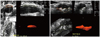
An increased IMT was defined as ≥ 1.0 mm in either or both carotid arteries, and the presence of an atherosclerotic plaque was defined as a focal structure that encroaches into the arterial lumen of at least 0.5 mm or 50% of the surrounding IMT value or demonstrates a thickness of > 1.5 mm as measured from the media-adventitial interface to the intima-lumen interface.18) The measurement plane was determined by scanning to find the largest plaque extension in longitudinal views of each plaque in the common, internal and external carotid arteries bilaterally.4-10) The image was then frozen, magnified, and the plaque was measured by tracing around the perimeter with a cursor on the screen. Fig. 1 shows the process of area determination by manual planimetry. Measurement of plaque area was acquired from tracing the plaque border by defining plaque as previously mentioned.4)7) The process was repeated until all plaques on both sides were measured. TPA was the sum of the areas of all plaques between the clavicle and angle of the jaw. Intra- and inter-observer intra-class correlations were 0.95 and 0.87, respectively.
Plaque volume measurement
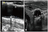
We measured plaque burden by 2D plaque area and 3D plaque volume (PV) simultaneously in selected cases, to evaluate the correlation between area and volume. Two observers were trained to identify and measure plaque volume with the 3D US images. Utilizing data from the 2D plaque area studies to localize plaques, 3D US images of plaque volumes were measured by manual planimetry utilizing the longitudinal scans of the CCA, bulb and internal carotid artery (ICA) (Fig. 2). Plaque volume calculations were carried out from the transverse plane starting at the edge of the plaque and moving along the longitudinal axis toward the opposite edge of the plaque. Plaque boundaries were traced using a mouse driven cross-haired cursor. Cross-sectional area of the plaque was delineated from 8-12 transverse sections of the image between the edges of the plaque, and the mean plaque volume value of 3 measurements of each plaque was used for analysis. This methodology for PV measurement and its validation has been described previously.19)20) Also, we calculated a modified plaque volume as the 2D US plaque area in the longitudinal plane multiplied by plaque width measured in the cross sectional plane. Fig. 3 shows the process of plaque determination by manual planimetry. Results of PV are presented as TPV from the 3D method and 2D PV from the 2D method. Intra- and inter-observer reliabilities were 0.93 and 0.94, respectively.
Coronary angiogram
An INTEGRIS BV 5000 (Philips Medical System, Best, Netherlands) was utilized to visualize the coronary artery stenosis. Quantitative measurements were analyzed by a workstation with dedicated software (WIN 32 version 3.3). Patients with at least one lesion > 50% within the main branches of the coronary arteries were considered to have significant CAD. Patients with minimal atherosclerotic lesions (≤ 50%) in the coronary arteries were not included. The association of mean IMT, TPA, and IMT with CAD severity was analyzed for patients with angiographically normal coronary arteries, one vessel CAD, two vessel CAD, and three vessel CAD.
Statistical analysis
Statistical analysis was performed with SPSS for Windows version 12.0 (SPSS Inc., Chicago, IL, USA). Results are presented as the mean ± standard deviation (SD) or percentage. For pairwise correlations between variables, we used data from all participants. Bland-Altman analysis was uses in comparison of data between 2D area-width volume and each matched 3D volume. Correlations between variables were made using Pearson correlation tests, and because of the large range of TPA and TPV results, analysis results using a logarithmic scale are reported in analysis of coronary angiography groups. Multivariate linear regression was used to determine sources of variation for transformed carotid US measurements, using selected continuous and discrete risk factor traits as covariates. Statistical significance was set at 0.05.
Results
Participants' clinical characteristics at baseline
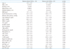
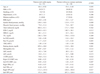
The mean age of the 107 study participants was 67.4 ± 9.8 years, and 60% (64/107) were male. In the entire sample, IMT, TPA, and TPV were 0.90 ± 0.26 mm, 0.42 ± 0.39 cm2, and 237.0 ± 301.2 mm3, respectively. Clinical characteristics and carotid artery parameters were analyzed according to the presence or absence of CAD, and are shown in Table 1. As expected, patients with CAD (n = 87) showed significantly increased IMT, TPA, and TPV bilaterally (all p < 0.05) (Table 1). When we dichotomized patients with CAD into stable angina or ACS, patients with ACS were more often male or smokers, with a higher prevalence of DM, lower HDL-C, and increased IMT, TPA, and TPV bilaterally (all p < 0.05) (Table 2).
Correlations among IMT, TPA, and TPV
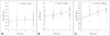
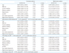
We found significant correlations for mean IMT : TPA, mean IMT : TPV and mean TPA : TPV of 0.448, 0.587, and 0.873, respectively (all p < 0.005) (Table 3). For the IMT : TPA pairs, r was 0.521 and 0.472 in right and left pairs respectively (all p < 0.001). For the IMT : TPV pair, r was 0.560 (p = 0.002) and 0.536 (p = 0.003) in right and left pairs respectively. For the TPA : TPV pair, r was 0.841 and 0.905 in right and left pairs respectively (all p < 0.001). Thus, the variables were significantly correlated, but the correlation coefficients were somewhat smaller for the comparisons of IMT and either TPA or TPV than the correlation coefficient for the TPA and TPV comparisons. Although there was no significant association of IMT and the severity of CAD (group 0 having normal coronary arteries, group 1 with one vessel CAD, group 2 with two vessel CAD, and group 3 with three vessel CAD), logarithmic transformation of TPA (Log TPA) and TPV (Log TPV) showed significant positive correlation with CAD severity (r = 0.436, p < 0.0003 for Log TPA and r = 0.593, p < 0.0001 for Log TPV) (Fig. 4).
Multivariate linear regression analysis
Multivariate linear regression was performed to examine the independent factors affecting the various parameters of carotid atherosclerosis (Table 4). Traditional cardiovascular risk factor such as age, sex, hypertension history, current smoking status, diabetes history, lipid profile and hs-CRP were examined as independent variables. Age was the only significant contributor to all three variables. However, there were differences in the associations of these measures with the remaining cardiovascular risk factors. Mean IMT was significantly associated only with hypertension, and not with current smoking status, DM, hs-CRP, or cholesterol in the study participants. In contrast, TPA was significantly associated with male sex, hypertension and LDL, and not with current smoking status, DM or hs-CRP in this sample. Finally, TPV was significantly associated with male sex, hs-CRP, and LDL-C, and not with hypertension, current smoking status, DM, hypertension or HDL-C in these patients.
Association between plaque volume by 2D and 3D US
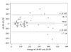
The 2D area-width volume were acquired from 100 plaques in 42 study patients with CCA plaques due to technical limitation in assessment of plaques in the carotid bulb or ICA in the remaining 65 patients. When we compare 2D area-width volume (110.2 ± 111.3 mm3) with each matched 3D volume (130.2 ± 101.4 mm3) by Bland-Altman analysis, the differences were within mean ± 1.96 SD, and the two methods may be used interchangeably (Fig. 5).
Discussion
In this study of atherosclerosis in patients with CAD, we simultaneously measured IMT, TPA, and TPV by US. We found that patients with ACS were more likely male or smokers, with a higher prevalence of DM, lower HDL-C, increased IMT, TPA, and TPV bilaterally. Secondly, although there were significant correlations among these parameters, the association between IMT with either TPA or TPV was less correlated than TPA and TPV. Finally, associations with traditional cardiovascular risk factors differed substantially between IMT, TPA, and TPV. While each was significantly associated with age, IMT was only significantly associated with hypertension, while TPA was associated with male sex, hypertension, and LDL-C, and TPV was associated with male sex, hs-CRP, and LDL-C. These findings were similar to the results of Spence and Hegele,21) who suggest that the three different US-derived measurements of carotid artery morphology, while somewhat correlated, might represent distinct intermediate traits with unique determinants and risk factor associations. The measurement of IMT as a surrogate marker for atherosclerosis is common in clinical practice. However, its accuracy has been questioned by the fact that the main predictors of medial hypertrophy or CCA intimal thickening are age and hypertension, which do not necessarily reflect the atherosclerotic process.22) In contrast, carotid plaque has been shown to be more closely related to CAD and to predict coronary events better than IMT.7)23) This is likely the result of carotid plaques predominantly occurring at sites of nonlaminar turbulent flow such as in the carotid bulb and the proximal ICA, but rarely in the CCA except in advanced atherosclerotic disease.24) As a measurement, IMT has the benefit of standardized acquisition, but the rigorous standards for the appropriate anatomical site interrogated to derive this measurement may also exclude some important information about the atherosclerotic burden in the remainder of the carotid arterial bed. Thus, a thorough scan of all carotid arteries, including plaque assessment, may increase sensitivity for identifying subclinical vascular disease.
According to our results, IMT was only significantly associated with age and hypertension, confirming that IMT mainly represents hypertensive medial hypertrophy, or thickening of smooth muscles in the media.7) In contrast, TPA was significantly associated with age, male sex, hypertension, and LDL-C, and TPV was significantly associated with age, male sex, hs-CRP and LDL-C. This is likely due to carotid plaques representing a later stage of atherogenesis related to inflammation, endothelial dysfunction, oxidative stress, and smooth muscle cell proliferation.21) Since age-related thickening of intimal and medial layers of CCA also occurs in the absence of overt atherosclerosis, IMT is not really atherosclerosis, but instead represents an indicator for cardiovascular risk. In contrast, carotid plaques are a distinctive phenotype of atherosclerosis, not a simple continuum of IMT progression, and they predict cardiovascular disease better than IMT.25) A recent meta-analysis provides further evidence that carotid plaque measurements are more strongly predictive of cardiovascular events than measurement of IMT.5)
However, the quantitative measurement of plaques is not standard practice for most Korean cardiologists, and the typical plaque description includes the number of individual plaques, the plaque thickness, and characterization of surrounding tissue, such as calcifications or various patterns of echogenicity. Accordingly, we tried to measure plaque burden as TPA and TPV according to the validated method in patients with well-established cardiovascular risk factors. While the simple correlations between IMT and plaque measurements were highly and statistically significant, the r-values between 0.4 and 0.6 indicated correlations that were only moderate. Conversely, there was a strong correlation between plaque measurement and both TPA and TPV, as large plaque measurements contribute to a large TPA and TPV, which is not always associated with increased IMT. Similarly, if there was only a long, slender plaque overlying the region used for IMT determination, the absence of other plaques contributed to low TPA and TPV for this individual. Thus, IMT does not always reflect total carotid disease burden. Additionally, we empirically measured plaque volume by the area-width method and compared these parameters with plaque volume by 3D US, which were shown to have significant positive correlations. However, there seems to be large discrepancies in the values over 200 mm3 in Bland-Altman plot analysis, we think there would be some potential role for this calculation in the assessment of plaque volume in limited cases with CCA plaques.
Of course, we recognize that our study is somewhat limited by several factors, including a small sample size and a unique study sample from which the findings may not be generalizable. As this was the first attempt to measure plaque area or volume in Korea, there would understandably be some technical limitations to plaque measurement. In our study, the golden standard of TPV was 3D US measurement and from our experience, 3D assessment of PV in ICA plaque was more difficult than CCA plaque because of angle, however, it was possible and feasibility of 3D PV is about 70-80%. However, for the assessment of 2D area-width PV, because we cannot achieve short axis view of ICA and bulb, ICA/bulb plaque could not be measured by this method, and feasibility of 2D PV is about 40-50%. So, it may have resulted in another level of imprecision. Moreover, it was difficult to define plaque border in cases with diffuse plaques connected by increased IMT. In these cases, we tried to measure the plaque border by defining plaque as suggested by Spence et al.4)7) Finally, we did not check TPV by another validated modality such as carotid CT angiography or magnetic resonance imaging. Despite this, we are confident that measurement of TPV by US would be more precise than the standard technique for measuring plaque thickness.
In conclusion, our results indicate that although there were significant correlations among the various ultrasound measures of carotid artery morphology, there seemed to be different biological determinants on IMT, TPA, and TPV. This has implications for studies of determinants of atherosclerosis that utilize indirect surrogate markers determined noninvasively. We might need to be selective about the particular measurements for specific applications.


 XML Download