

 PDF
PDF ePub
ePub Citation
 Print
Print


Introduction
Right ventricular (RV) pressure overload induces biventricular distortion and makes it difficult to assess biventricular systolic function by conventional 2-dimensional echocardiography.1) There are few studies in which real-time 3-dimensional echocardiography (RT3DE) is used for the assessment of RV and left ventricular (LV) volumes and functions of patients with biventricular distortion due to cor pulmonale.1-6) Cardiac magnetic resonance (CMR) imaging is considered the gold standard for noninvasive measurement of cardiac chamber volume owing to high levels of accuracy and reproducibility in the measurement of biventricular volume.7)8) However, CMR imaging has limitations in evaluating patients with severe dyspnea who cannot tolerate long examination time. It has been demonstrated that 64-slice multi-detector cardiac computed tomography (64-MDCT) allows for accurate assessment of biventricular function and has good correlation with CMR results in patients who are not capable of holding their breath for extended periods.9)10) RT3DE also has good correlations with CMR imaging results in the measurement of right and left ventricular volumes and ejection fractions (EFs).11-16) This is a validation study for ventricular volume measurement by RT3DE in patients with biventricular distortion due to cor pulmonale using to 64-MDCT as a reference method.
Methods
Study subjects
We prospectively recruited 30 consecutive adult patients who showed flattening or reverse curvature of the interventricular septum and severe pulmonary hypertension due to cor pulmonale on conventional echocardiography between December 2009 and January 2011. As a part of our protocol, 26 patients also underwent 64-MDCT and RT3DE on the same day in order to minimize confounding variables for volumes in the right and left ventricular assessment (time delay between 64-MDCT and RT3DE = 3.7 ± 2.1 hours). Four patients were excluded from RT3DE evaluation due to poor echo windows for analysis. Our study included a total of 22 patients (59.3 ± 16.6 years of age, 10 males) who showed biventricular distortion due to cor pulmonale [RV systolic pressure (RVSP) = 66.8 ± 19.7 mmHg].
Exclusion criteria were patients with atrial fibrillation, left bundle-branch block, heart failure caused by other conditions such as primary left heart failure, abnormal shunt flow, congenital anomalies, moderate or severe mitral or aortic valvular disease, pericardial effusion, renal dysfunction (creatinine > 1.3 mg/dL) and known allergy to iodine.
Written informed consents were obtained from all patients. The study protocol was approved by the Ethics Committee of Daejeon St. Mary's Hospital, College of Medicine, The Catholic University of Korea, Daejeon, Republic of Korea (DC10 OISI0031).
Echocardiographic examination
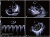
Two-dimensional transthoracic echocardiography was performed for all subjects. The severity of D-shaped deformation was assessed by LV eccentricity index (the ratio of diameters of perpendicular/parallel to the interventricular septum on parasternal short axis images of the papillary muscle level) (Fig. 1). To quantify the degree of RV dysfunction, fractional area change (FAC) and RVSP were measured according to the current guidelines.17) Inferior vena cava diameter ≤ 2.1 cm that collapses > 50% with a sniff suggests normal right atrial (RA) pressure of 3 mmHg (range, 0-5 mmHg), whereas inferior vena cava diameter diameter > 2.1 cm that collapses < 50% with a sniff suggests high RA pressure of 15 mmHg (range, 10-20 mmHg). Severe pulmonary hypertension was defined as RVSP ≥ 65 mmHg on transthoracic echocardiography.
Assessment of real-time 3D echocardiography
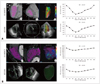
RT3DE was performed from the apical window using an Acuson SC2000 scanner (Siemens Medical Solutions Inc., Mountain View, CA, USA) equipped with a fully sampled matrix array 4Z1c transducer. Images were acquired by single beat full volume capture by experienced investigators who were blinded to 64-MDCT data and the frame rate was adjusted between 12 and 15 frames/s. Furthermore, images were analyzed with an embedded Volume Cardiac Analysis Package (Siemens Medical Solutions Inc.) on the Acuson SC2000 system that allowed for automated delineation (auto-contouring) of the endocardium of the LV and semi-automated contouring of the endocardium of the RV throughout the entire cardiac cycle (Fig. 2). Unlike the auto-contoured LV, the RV required additional steps for contouring; landmarks in tricuspid valve, mitral valve, and LV apex were first set (step 1) and RV contours for the 4 chambers, coronal and sagittal views were set (step 2). Then contour tracking was checked (step 3) and RV was analyzed (step 4). Some contours were corrected manually if needed.
Assessment of 64-slice multi-detector cardiac computed tomography
Patients underwent cardiac computed tomography using a 0.5 mm × 64-detector scanner (Sensation 64, Siemens Medical Solutions, Malvern, PA, USA). Because patients had severe dyspnea, β-blockers were not used. The mean heart rate was 72 ± 14 beat/min. 70 ± 5.6 mL of contrast agent (Ultravist 370, Bayer HealthCare, Berlin, Germany) was administered at a rate of 5 mL/s via an 18-gage venous access placed in the antecubital vein, followed by a 40 mL saline chaser at the same flow rate. Imaging studies were performed according to a retrospectively gated cardiac 64-MDCT protocol [volume/scan distance = 180 mm, time = 18 seconds, tube current = 770 mA, tube voltage = 120 kV, gantry rotation time = 0.33 sec (temporal resolution 165 ms), scanning field of view 350 mm, slice thickness = 0.5 mm]. Total radiation dose was between 10 and 16 mSv. The 64-MDCT images were analyzed using a custom cardiac software package (Aquarius Workstation, Terarecon Inc., San Mateo, CA, USA) by an experienced investigator who was blinded to echocardiographic data. LV and RV endocardial borders were determined by the Terarecon density threshold algorithm for each slice (Fig. 2). Using the retrospective electrocardiographic gating technique, the images were reconstructed from 20 phases of the cardiac cycle. Semi-automated endocardial border tracing allowed for time-volume analyses of both the LV and RV, such as end-diastole, end-systole, stroke volume and EF (Fig. 2).
Statistical analysis
Pearson's correlation coefficient was used for comparison. Bland-Altman analysis (MedCalc, version 8.2.1.0, MedCalc Software, Mariakerke, Belgium) was used to determine the limits of agreement between the end-diastolic volumes, end-systolic volumes and EFs of both right and left ventricles on 64-MDCT and RT3DE. Data is expressed as the mean ± standard deviation. Interobserver and intraobserver variability of RT3DE measurement were obtained from biventricular measurements from 9 randomly selected patients. The intraclass correlation coefficient and Wilcoxon's signed-rank test were used for interobserver and intraobserver variability. A p value of < 0.05 was considered statistically significant. Statistical analyses were performed by using the Statistical Package for Social Sciences for Windows (version 15.0, SPSS Inc., Chicago, IL, USA).
Results
Patient population
Patient characteristics are summarized in Table 1. A total of 22 patients (59.3 ± 16.6 years of age; 10 males and 12 females) with severe pulmonary hypertension (RVSP = 66.8 ± 19.7 mmHg) due to cor pulmonale were included in this study. All patients were categorized into either New York Heart Association (NYHA) functional class III or IV. The etiologies of cor pulmonale were chronic bronchitis, bronchiectasis and emphysema. The interventricular septum was shifted in all patients, resulting in D-shaped LV (LV eccentricity index = 1.87 ± 0.57).
RV and LV functions in patients with cor pulmonale
FAC as measured by 2-dimensional echocardiography and right ventricle ejection fraction (RVEF) as measured by RT3DE, were markedly decreased (FAC by 2-dimensional echocardiography, 22.0 ± 8.8%; RVEF by RT3DE, 27.0 ± 12.3%; RV stroke volume by RT3DE, 29.26 ± 16.11 mL). Left ventricle ejection fraction (LVEF), as measured by RT3DE, was relatively well preserved, but the LVs were underfilled and showed decreased stroke volumes (LVEF, 49.3 ± 7.13%; LV stroke volume, 29.16 ± 9.93 mL).
Correlation between RV function and LV eccentricity index
The severity of LV eccentricity index was not correlated with EF of the LV (r = -0.06, p = 0.77) but with EF of the RV as measured by RT3DE (r = 0.44, p = 0.03). Also, it was reversely correlated with RVSP (r = -0.43, p = 0.04).
Correlation and agreement between RT3DE and 64-MDCT
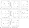
There were moderate degree agreements on Bland-Altman plots in LV end-diastolic and systolic volumes measured by RT3DE and 64-MDCT (Fig. 3A and B). Relatively low degree agreements are noted between Bland-Altman plots of the end-diastolic and systolic volumes of RV as measured by both methods (Fig. 3E and F). However, moderate degree of agreements are noted between Bland-Altman plots of the EFs and stroke volumes of the LV and RV as measured by both methods (Fig. 3C, D, G and H). LVEF was not measured by 2-dimensional echocardiography because of inadequate images on the 2-chamber view caused by the distorted and underfilled LV (Fig. 1B). There were moderate correlations between left and right ventricular volumes as measured by RT3DE and 64-MDCT except for LV end-systolic volume (Table 2).
Interobserver and intraobserver variability of RT3DE measurements of biventricular volumes
Interobserver and intraobserver variability are summarized in Table 3. The intraclass correlation coefficients ranged from 0.84 to 0.96 between observers and from 0.86 to 0.94 between measurements.
Discussion
Enrolled patients had the following typical signs of cor pulmonale. There were 1) right ventricular and atrial enlargement with a reduced left-ventricular cavity and 2) reversal of septal curvature.1-6) Clinically, echocardiography is an important diagnostic modality because it is noninvasive and easily available for patients with severe dyspnea. Also, it allows for the assessment of RV dilatation, cardiac function and pulmonary artery pressure.1-6) However, the window view is occasionally insufficient because of hyperinflated lungs, especially in patients with emphysema. In our study, 13.5% of enrolled patients had emphysematous lung disease.
Feasibility of RT3DE and 64-MDCT in the assessment of biventricular distortion due to cor pulmonale
Numerous studies have shown that RT3DE is a feasible method for measuring LV and RV volumes and is more accurate than 2-dimensional approaches.18-20) There was severe apex distortion in most of 2 chamber views obtained by 2-dimensional approaches, as shown in Fig. 1B, since the subjects of this study had flattening or reverse curvature of the interventricular septum. Therefore, LV volumes could not be accurately measured with Simpson's rule. However, we were able to obtain volume data using RT3DE although it had modest correlation with 64-MDCT. This showed that RT3DE could supplement the limitations of 2-dimensional echocardiography. However, 12-15 volume rates of RT3DE was lower than 20 phase of 64-MDCT. Furthermore, this study included patients with relatively severe dyspnea (NYHA III-IV) and several unstable patients, resulting in the underrated feasibility. LV end-systolic volumes were also significantly reduced in underfilled LVs inducing a greater error range and border delineation was also difficult in distorted and underfilled LVs. Owing to these factors, the statistical significance of the correlation between RT3DE and 64-MDCT was thought to be reduced. Moreover, although 64-MDCT provides accurate information on biventricular function with good correlation with CMR in patients with pulmonary hypertension who are unsuitable for CMR,9)21) 64-MDCT is not a gold standard tool for volume measurement as Sugeng et al.22) have demonstrated that 64-MDCT tends to overestimate LV volumes compared to CMR values.
With regards of RV volume, it was reported that the volumes and EF of a dilated RV may be underestimated with 3DE, which may be explained by the disability of 3DE to visualize the entire RV apex in some excessively dilated RV.15-17) Also, there were relatively low degree agreements between Bland-Altman plots of the end-diastolic and systolic volumes of RV as measured by RT3DE and 64-MDCT in present study. However, stroke volume of RV with measured RT3DE had moderate degree agreements with measured 64-MDCT and end-diastolic and end-systolic volume of RV had moderate correlations.
RV and LV volumes and functions measured by RT3DE in patients with cor pulmonale
The evaluation of RV function is important for assessing prognosis in severe pulmonary hypertension associated with chronic obstructive lung disease. The RV is considered a "volume" rather than a "pressure" pump.3)23) When pulmonary hypertension progresses, the RV dilates in both end-diastolic and end-systolic volumes. Cor pulmonale leads to changes in RV and LV structures or functions caused by pulmonary hypertension associated with the involvement of the lung or its vasculature.2-6)24-26) There are few studies in which cardiac volumes and functions of patient with cor pulmonale are assessed by RT3DE. In our study, FAC measured by 2-dimensional echocardiography and RVEF measured by RT3DE were significantly decreased.
LVEF was preserved, but LV stroke volume was decreased as with RV stroke volume. This implies that hemodynamically unstable cor pulmonale may be attributed to the underfilled LV along with decreased LV systolic and diastolic volumes, but not to the efficacy of LV wall motion. Therefore, accurate assessment of stroke volumes may be vital in patients with unstable vital signs such as cor pulmonale. RT3DE may be considered to be a modest method for these patients. In addition, it may be beneficial to evaluate hemodynamic status of patients in whom CMR or cardiac catheterization is not feasible. Furthermore, the eccentricity index of the LV, which can be easily measured by 2-dimensional echo, may be also considered to be a useful parameter because it indirectly provides information on RV function and RVSP.
This study has some limitations. First limitation is the small sample size. Another limitation is the relatively low temporal resolutions of 64-MDCT and RT3DE due to the hyperinflation of the lungs and avoidance of β-blockers. Cardiac catheterization was not feasible to evaluate hemodynamic parameters of the subjects in this study. Contrast agents which might have affected RV and LV volumes were only used during 64-MDCT but not during RT3DE. 64-MDCT has the advantage of more rapid image acquisition, which is a benefit to patients with cor pulmonale. However, since 64-MDCT is not considered the gold standard in this respect, validation of a third technique such as RT3DE should be performed against the "primary" modality such as CMR. The subjects of this study were most unlikely to be able to tolerate CMR due to short of breath.
In conclusion, this study results suggest that RT3DE may be a modest method to evaluate stroke volume comparing with 64-MDCT.


 XML Download