Journal List > J Cardiovasc Ultrasound > v.21(2) > 1018274

Moustafa, Ejaz, Momenah, AlKhaldi, Zuhairy, AlMoukirish, and Mookadam: Unusual Case of Cor Triatriatum Sinister
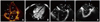
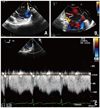
A 26-year-old man was evaluated for progressive shortness of breath on mild exertion. Physical examination was unremarkable. The electrocardiogram revealed left atrial (LA) enlargement. Transthoracic echocardiography (TTE) and cardiovascular magnetic resonance (CMR) demonstrated normal biventricular size and systolic function with severely dilated LA and no significant valvulopathy. A thin mobile complex septated membrane was noted within the LA with no clear fenestrations (Fig. 1A and B, Supplementary movie 1 and 2). Transesophageal echocardiogram (TEE) revealed that the membrane is attached to the lateral wall at the coumadin ridge with color turbulence at the site of attachment yet no fenestrations were seen. Medially, the membrane was attached to the inter-atrial septum just above the fossa ovalis with possible multiple fenestrations by color Doppler. The calculated peak/mean gradients were 13-14/9-10 mmHg respectively at both ends. A septum within the membrane was visualized extending from its lateral wall and attached to the inter-atrial septum just above the fossa ovalis (Fig. 1C, D and 2, Supplementary movie 3-7). A decision was made to proceed with surgical correction. The findings of the TTE/TEE/CMR were confirmed and the atrial membrane was excised around its periphery. Recovery from the surgery was uneventful and he was asymptomatic 6 months afterwards. Echocardiography revealed neither residual membrane nor stenosis of the pulmonary veins.
Cor triatriatum sinister is a rare congenital anomaly wherein the LA is subdivided by a fibromuscular membrane into two chambers. It usually manifests early in life, nonetheless, it might remain asymptomatic till adulthood. Clinical scenario can imitate that of mitral stenosis due to the obstructive nature of the fenestrations. Surgical resection of the membrane is the treatment of choice for symptomatic patients.1-3)

Figures and Tables

Fig. 1
Transthoracic echocardiography apical 4-chamber view revealing a complex membrane (arrow) with its septum (arrowhead) subdividing the LA into two chambers (A). Cardiovascular magnetic resonance 4-chamber steady-state free precession image showing the membrane (arrow) with its septum (arrowhead) (B). Transesophageal echocardiogram mid-esophageal 4-chamber (C) and modified 2-chamber (D) views demonstrating the membrane which is attached to the coumadin ridge just below the left pulmonary veins (D). LA: true left atrium, LV: left ventricle, PVC: pulmonary venous chamber, RA: right atrium, RV: right ventricle.
jcu-21-100-g001
Fig. 2
Transesophageal echocardiogram modified mid-esophageal views showing the lateral (A) and medial (B) attachments of the membrane with color turbulence and possible multiple fenestrations (B, arrows). Continuous wave Doppler through the lateral edge of the membrane showing significant gradient across, both systolic and diastolic (C). PVC: pulmonary venous chamber.
jcu-21-100-g002

References

1. Church WS. Congenital malformation of the heart: abnormal septum in the left auricle. Trans Pathol Soc Lond. 1868; 19:188–190.
2. van Son JA, Danielson GK, Schaff HV, Puga FJ, Seward JB, Hagler DJ, Mair DD. Cor triatriatum: diagnosis, operative approach, and late results. Mayo Clin Proc. 1993; 68:854–859.
crossref
3. Alphonso N, Nørgaard MA, Newcomb A, d'Udekem Y, Brizard CP, Cochrane A. Cor triatriatum: presentation, diagnosis and long-term surgical results. Ann Thorac Surg. 2005; 80:1666–1671.
crossref

Supplementary movie legends

Movie 1 and 2

Transthoracic echocardiography parasternal long axis (1) and 4-chamber (2) views revealing a complex mobile membrane with its septum.
Movie 1
Movie 2

Movie 3, 4 and 5

Transesophageal echocardiogram midesophageal 4-chamber (3), apical long axis (4) and modified 2-chamber (5) views demonstrating the complex membrane. Medially, the membrane, with its septum, is attached to the inter-atrial septum just above the fossa ovalis (3) and laterally is attached to the coumadin ridge just below the left pulmonary veins (5).
Movie 3
Movie 4
Movie 5

Movie 6 and 7

Transesophageal echocardiogram modified mid-esophageal views showing the lateral (6) and medial (7) attachments of the membrane with color turbulence and possible multiple fenestrations (7).
Movie 6
Movie 7
TOOLS
Similar articles