

 PDF
PDF ePub
ePub Citation
 Print
Print


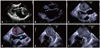
A 74-year-old Japanese female was diagnosed as chronic atrial fibrillation, and anticoagulant therapy with warfarin started. One year after anticoagulant therapy, she was referred to our center for evaluation of cardiac function. Transthoracic echocardiography by Philips iE33 ultrasound system with S5-1 transducer (Philips Medical Systems, Andover, MA, USA) revealed huge left atrial thrombus (53 × 36 mm) (Fig. 1A). Left ventricular ejection fraction was 44% with no focal asynergy. Her blood coagulation study revealed no significant problems. At that time, her prothrombin time-international normalized ratio (PT-INR) was 1.7, therefore we adjusted warfarin dose to maintain PT-INR levels at 2.0-3.0. Several weeks later, we performed transthoracic echocardiography by Siemens ACUSON SC2000 ultrasound system with 4V1 transducer (Siemens Ultrasound, Mountain View, CA, USA) and found the huge thrombus had almost disappeared (Fig. 1B). Trans-esophageal echocardiography by ACUSON SC2000 ultrasound system with V5M trans-esophageal transducer (Siemens Ultrasound, Mountain View, CA, USA) also revealed no thrombus in the left atrium and revealed severe spontaneous echo contrast (Fig. 1C-F). Left atrial appendage flow velocity was about 10 cm/sec. Black defects of spontaneous echo contrast are completely same region shown as mild mitral regurgitation in color Doppler imaging (Fig. 1D vs. C, F vs. E, and Supplementary movie 1).
This patient showed severe spontaneous echo contrast in her left atrium. Spontaneous echo contrast seemed to be due to atrial fibrillation, reduced left ventricular function, and large left atrium.1) Furthermore, there was only mild mitral regurgitation. Previous reports say that mitral regurgitation has protective effect on left atrial blood stasis. But the effect is limited to patients with severe mitral regurgitation.2) Previous reports have already mentioned this protective effect of severe mitral regurgitation against stroke.3)4) Especially in patients with atrial fibrillation, severe mitral regurgitation can disperse the aggregation of blood component in left atrium, which resulted in the lower risk of stroke than patients without severe mitral regurgitation. Furthermore, Kranidis et al.5) reported that when the mitral regurgitation jet volume-to-left atrial volume ratio is small, the regurgitant-stirring effect is reduced, and blood stasis in the left atrium is increased. Although this is important discussion, there was no direct evidence to show this mechanism. Severe spontaneous echo contrast could visualize mitral regurgitation clearly without using color Doppler flow imaging (Fig. 1C, E) in this case. In other words, black defects in spontaneous echo contrast meant that the mitral regurgitation disrupted left atrial blood stasis. This echocardiographic imaging supports the previous hypothesis that severe mitral regurgitation can reduce left atrial blood stasis, which results in lower incidence of spontaneous echo contrast. This imaging is important, because black defects of spontaneous echo contrast support understanding the protective aspect of mitral regurgitation especially against spontaneous echo contrast visually.

 XML Download