

 PDF
PDF ePub
ePub Citation
 Print
Print


Introduction
Patent foramen ovale (PFO) is a slit or tunnel like passage in the interatrial septum formed by failure of postnatal fusion of the septum primum and septum secundum. Persistent PFO occurs in around 20-25% of the adult population.1) PFO has been known to be the cause of transient ischemic attacks (TIA) or stroke. Meta-analyses and observational studies indicate that the prevalence of PFO is approximately 3-fold higher in patients with cryptogenic stroke and migraine compared to controls. Conversely, observational evidences indicate a 2-3-fold increased prevalence of migraine and cerebrovascular events in PFO carriers.2) Transcatheter device closure has been the treatment of choice for these defects.
The persistent left superior vena cava (LSVC) is the most common abnormality of systemic venous return. It has been observed in 0.5% of the general population and in up to 10% of patients with congenital heart disease.3)4) This anomaly is due to an abnormal development of the sinus venosus in the early stages of fetal life. In 92% of cases, drainage occurs in the right atrium; in the remainder of cases, drainage occurs in the left atrium (LA), either directly or through an unroofed coronary sinus.5) Combined defect of abnormal drainage of LSVC to left superior pulmonary vein (LSPV) in PFO patients is uncommon, and we report simultaneous device closure of LSVC with Amplatzer® Vascular Plug II (St. Jude Medical, St. Paul, MN, USA).
Case
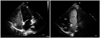
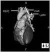
A 37-year-old female patient with chief complaint of left arm weakness (duration less than 1 minute) and left-sided lip twitching was diagnosed with PFO under evaluation at Myongji Hospital. Laboratory test results showed normal value of erythrocyte sedimentation rate, C-reactive protein, no abnormality of coagulopathy, and no suspicious sign of vasculitis. While continuation of warfarin treatment and medication in regards of rhematoid arthritis diagnosed in 2007, she was referred to Pediatrics Cardiology for PFO closure. Brain magnetic resonance imaging showed signs of stroke and transcatheter closure of PFO was scheduled. Contrast echocardiography performed via the left upper extremity prior to the procedure showed sequential filling of the bubble in the LA followed by left ventricle and then through the PFO to the right atrium (Fig. 1). For further evaluation, heart computed tomography (CT) was performed and an abnormal connection of LSVC to LSPV which drained into LA was diagnosed (Fig. 2).
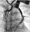
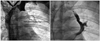
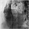
Under local anesthesia, we first evaluated the patient's hemodynamic data to confirm any presence of pulmonary hypertension and patency of coronary artery which showed normal findings. Through venous approach via right femoral vein, 7 French guiding catheter passed right atrium and innominate vein approaching to LSVC. The proximal and distal part of the LSVC were measured 9.8 and 9.6 mm, respectively by angiography (Fig. 3). The abnormal connection between LSVC and LSPV was closed using the Amplatzer® Vascular Plug II (diameter = 12 mm; St. Jude Medical, St. Paul, MN, USA) (Fig. 4). After checked up size of PFO by sizing balloon, transcatheter PFO closure with Amplatzer® PFO occluder (diameter = 25 mm; St. Jude Medical, St. Paul, MN, USA) was also performed (Fig. 5). Follow-up echocardiogram showed complete occlusion of flow through the LSVC and PFO.
Discussion
In recent years, transthoracic echocardiogram (TTE) or transesophageal echocardiography in patients who have had TIA or stroke has become a routine assessment. A contrast echocardiography is usually performed by injecting microbubbles through the peripheral intravenous (IV) line, and acted the Valsalva maneuver to determine the presence of right-to-left shunting across the PFO. In our patient, the IV line was first started on the right arm and the contrast echocardiography was performed, which proved the presence of PFO. On contrast echo conducted on Rt. arm, the enhancement was seen at not only Rt. side of heart, but also Lt. side. On careful review by an experienced doctor, small amount of direct flow into the LA was recognized. Another IV line was started on the left arm of the patient. The contrast echo was done as a same way on Lt. arm. On repeated contrast echocardiography, massive amount of microbubbles draining straight to LA was detected. Heart CT was performed to determine the precise LSVC drainage, resulting persistent LSVC persistent LSVC connection to LSPV was diagnosed.
Incidence of persistent LSVC is 0.5% of the general population and increased to 4.3% in patients with congenital heart disease.6)7) Persistent LSVC draining into the coronary sinus and the right atrium is of no hemodynamic significance. But persistent LSVC draining directly or through an unroofed coronary sinus into the left atrium represents the same risks as all other lesions with right-to-left shunt and forms a substrate for paradoxical embolism. If complications related to the right-to-left shunt occur, correction is indicated.8) In this case of our patient, relatively young age at the time of stroke development probably indicates that both PFO and persistent LSVC was the cause of paradoxical embolism.
Although our team came across the diagnosis by the keen sense of an experienced doctor, in order to increase the chance of the detection of persistent LSVC, the authors recommend performing contrast echocardiography through the left arm peripheral IV line, or through both right and left, if the patients consent to it. However, the right-to-left shunt through this persistent vein is usually not sufficient enough to provoke clinically significant systemic desaturation. Therefore, if clinically suspected, a complete evaluation for this anomaly should also be considered.
There are many devices used to occlude vessels, the Amplatzer® vascular device is easily conformable and has a wide range of sizes.9) After device closure, the patient was followed-up regularly thereafter and she is free from additional neurologic attacks, and on follow-up TTE, no residual shunting through both device was detected.
In conclusion, we report a successful simultaneous closure of PFO using the Amplatzer® PFO occluder (St. Jude Medical, St. Paul, MN, USA) and persistent LSVC connection to LSPV using the Amplatzer® Vascular Plug II (St. Jude Medical, St. Paul, MN, USA). In routine work-up performed on TIA or stroke patients, in order to increase the chance of diagnosing additional right-to-left shunting other than PFO, the authors suggest performing contrast echocardiography through the left arm peripheral IV line, or on both arm if the patients consent to it.10)
 XML Download